1RD3

2.5A Structure of Anticoagulant Thrombin Variant E217K


Experimental Data Snapshot

  • Method: X-RAY DIFFRACTION
  • Resolution: 2.50 Å
  • R-Value Free: 0.259 
  • R-Value Work: 0.229 

wwPDB Validation   3D Report Full Report


This is version 2.2 of the entry. See complete history


Literature

Crystal Structure of Anticoagulant Thrombin Variant E217K Provides Insights into Thrombin Allostery

Carter, W.J.Myles, T.Gibbs, C.S.Leung, L.L.Huntington, J.A.

(2004) J Biol Chem 279: 26387-26394

  • DOI: https://doi.org/10.1074/jbc.M402364200
  • Primary Citation of Related Structures:  
    1RD3

  • PubMed Abstract: 

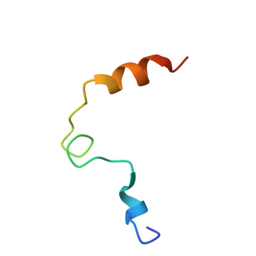
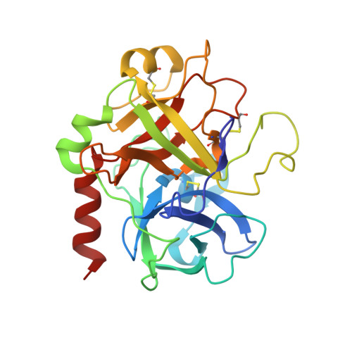
    Thrombin is the ultimate protease of the blood clotting cascade and plays a major role in its own regulation. The ability of thrombin to exhibit both pro- and anti-coagulant properties has spawned efforts to turn thrombin into an anticoagulant for therapeutic purposes. This quest culminated in the identification of the E217K variant through scanning and saturation mutagenesis. The antithrombotic properties of E217K thrombin are derived from its inability to convert fibrinogen to a fibrin clot while maintaining its thrombomodulin-dependent ability to activate the anticoagulant protein C pathway. Here we describe the 2.5-A crystal structure of human E217K thrombin, which displays a dramatic restructuring of the geometry of the active site. Of particular interest is the repositioning of Glu-192, which hydrogen bonds to the catalytic Ser-195 and which results in the complete occlusion of the active site and the destruction of the oxyanion hole. Substrate binding pockets are further blocked by residues previously implicated in thrombin allostery. We have concluded that the E217K mutation causes the allosteric inactivation of thrombin by destabilizing the Na(+) binding site and that the structure thus may represent the Na(+)-free, catalytically inert "slow" form.


  • Organizational Affiliation

    University of Cambridge, Department of Haematology, Division of Structural Medicine, Thrombosis Research Unit, Cambridge Institute for Medical Research, Wellcome Trust/MRC Building, Hills Road, Cambridge CB2 2XY, United Kingdom.


Macromolecules
Find similar proteins by:  (by identity cutoff)  |  3D Structure
Entity ID: 1
MoleculeChains Sequence LengthOrganismDetailsImage
Prothrombin
A, C
36Homo sapiensMutation(s): 0 
Gene Names: F2
EC: 3.4.21.5
UniProt & NIH Common Fund Data Resources
Find proteins for P00734 (Homo sapiens)
Explore P00734 
Go to UniProtKB:  P00734
PHAROS:  P00734
GTEx:  ENSG00000180210 
Entity Groups  
Sequence Clusters30% Identity50% Identity70% Identity90% Identity95% Identity100% Identity
UniProt GroupP00734
Sequence Annotations
Expand
  • Reference Sequence
Find similar proteins by:  (by identity cutoff)  |  3D Structure
Entity ID: 2
MoleculeChains Sequence LengthOrganismDetailsImage
Prothrombin
B, D
259Homo sapiensMutation(s): 1 
Gene Names: F2
EC: 3.4.21.5
UniProt & NIH Common Fund Data Resources
Find proteins for P00734 (Homo sapiens)
Explore P00734 
Go to UniProtKB:  P00734
PHAROS:  P00734
GTEx:  ENSG00000180210 
Entity Groups  
Sequence Clusters30% Identity50% Identity70% Identity90% Identity95% Identity100% Identity
UniProt GroupP00734
Sequence Annotations
Expand
  • Reference Sequence
Oligosaccharides

Help

Entity ID: 3
MoleculeChains Length2D Diagram Glycosylation3D Interactions
alpha-D-mannopyranose-(1-6)-beta-D-mannopyranose-(1-4)-2-acetamido-2-deoxy-alpha-D-glucopyranose-(1-4)-[alpha-L-fucopyranose-(1-6)]2-acetamido-2-deoxy-beta-D-glucopyranose
E
5N-Glycosylation
Glycosylation Resources
GlyTouCan:  G89753YM
GlyCosmos:  G89753YM
GlyGen:  G89753YM
Entity ID: 4
MoleculeChains Length2D Diagram Glycosylation3D Interactions
2-acetamido-2-deoxy-alpha-D-glucopyranose-(1-4)-[alpha-L-fucopyranose-(1-6)]2-acetamido-2-deoxy-beta-D-glucopyranose
F
3N-Glycosylation
Glycosylation Resources
GlyTouCan:  G19870TQ
GlyCosmos:  G19870TQ
GlyGen:  G19870TQ
Experimental Data & Validation

Experimental Data

  • Method: X-RAY DIFFRACTION
  • Resolution: 2.50 Å
  • R-Value Free: 0.259 
  • R-Value Work: 0.229 
  • Space Group: P 21 21 21
Unit Cell:
Length ( Å )Angle ( ˚ )
a = 73.23α = 90
b = 107.62β = 90
c = 136.03γ = 90
Software Package:
Software NamePurpose
MOSFLMdata reduction
SCALAdata scaling
MOLREPphasing
CNSrefinement
CCP4data scaling

Structure Validation

View Full Validation Report


Entry History 

Deposition Data

Revision History  (Full details and data files)

  • Version 1.0: 2004-05-04
    Type: Initial release
  • Version 1.1: 2008-04-29
    Changes: Version format compliance
  • Version 1.2: 2011-07-13
    Changes: Derived calculations, Version format compliance
  • Version 2.0: 2020-07-29
    Type: Remediation
    Reason: Carbohydrate remediation
    Changes: Advisory, Atomic model, Data collection, Derived calculations, Structure summary
  • Version 2.1: 2021-10-27
    Changes: Database references, Structure summary
  • Version 2.2: 2023-08-23
    Changes: Data collection, Refinement description