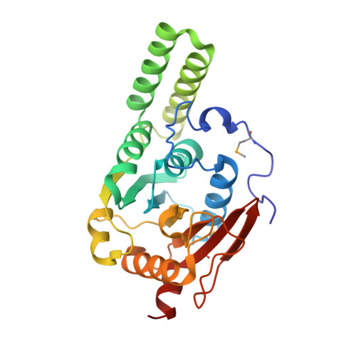
Crystal Structure of the Avilamycin Resistance-Conferring Methyltransferase Avira from Streptomyces Viridochromogenes
Mosbacher, T.G., Bechthold, A., Schulz, G.E.(2003) J Mol Biol 329: 147
- PubMed: 12742024
- DOI: https://doi.org/10.1016/s0022-2836(03)00407-8
- Primary Citation of Related Structures:
1O9G, 1O9H - PubMed Abstract:
The emergence of antibiotic-resistant bacterial strains is a widespread problem in contemporary medical practice and drug design. It is therefore important to elucidate the underlying mechanism in each case. The methyltransferase AviRa from Streptomyces viridochromogenes mediates resistance to the antibiotic avilamycin, which is closely related to evernimicin, an oligosaccharide antibiotic that has been used in medical studies. The structure of AviRa was determined by X-ray diffraction at 1.5A resolution. Phases were obtained from one selenomethionine residue introduced by site-directed mutagenesis. The chain-fold is similar to that of most methyltransferases, although AviRa contains two additional helices as a specific feature. A putative-binding site for the cofactor S-adenosyl-L-methionine was derived from homologous structures. It agrees with the conserved pattern of interacting amino acid residues. AviRa methylates a specific guanine base within the peptidyltransferase loop of the 23S ribosomal RNA. Guided by the target, the enzyme was docked to the cognate ribosomal surface, where it fit well into a deep cleft without contacting any ribosomal protein. The two additional alpha-helices of AviRa filled a depression in the surface. Since the transferred methyl group of the cofactor is in a pocket beneath the enzyme surface, the targeted guanine base has to flip out for methylation.
Organizational Affiliation:
Institut für Organische Chemie und Biochemie, Albert-Ludwigs-Universität, Albertstr. 21, Freiburg im Breisgau, 79104, Germany.