1O7S

High resolution structure of Siglec-7


Experimental Data Snapshot

  • Method: X-RAY DIFFRACTION
  • Resolution: 1.75 Å
  • R-Value Free: 0.248 
  • R-Value Work: 0.221 
  • R-Value Observed: 0.221 

wwPDB Validation   3D Report Full Report


Ligand Structure Quality Assessment 


This is version 2.2 of the entry. See complete history


Literature

High Resolution Structures of Siglec-7 - Insights Into Ligand Specificity in the Siglec Family

Alphey, M.S.Attrill, H.Crocker, P.R.Van Aalten, D.M.F.

(2003) J Biol Chem 278: 3372

  • DOI: https://doi.org/10.1074/jbc.M210602200
  • Primary Citation of Related Structures:  
    1O7S

  • PubMed Abstract: 

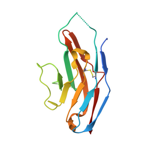
    Sialic acid-binding immunoglobulin-like lectins (Siglecs) recognize sialylated glycoconjugates and play a role in cell-cell recognition. Siglec-7 is expressed on natural killer cells and displays unique ligand binding properties different from other members of the Siglec family. Here we describe the high resolution structures of the N-terminal V-set Ig-like domain of Siglec-7 in two crystal forms, at 1.75 and 1.9 A. The latter crystal form reveals the full structure of this domain and allows us to speculate on the differential ligand binding properties displayed by members of the Siglec family. A fully ordered N-linked glycan is observed, tethered by tight interactions with symmetry-related protein molecules in the crystal. Comparison of the structure with that of sialoadhesin and a model of Siglec-9 shows that the unique preference of Siglec-7 for alpha(2,8)-linked disialic acid is likely to reside in the C-C' loop, which is variable in the Siglec family. In the Siglec-7 structure, the ligand-binding pocket is occupied by a loop of a symmetry-related molecule, mimicking the interactions with sialic acid.


  • Organizational Affiliation

    Division of Cell Biology and Immunology, School of Life Sciences, University of Dundee, Dundee DD1 5EH, Scotland, United Kingdom.


Macromolecules
Find similar proteins by:  (by identity cutoff)  |  3D Structure
Entity ID: 1
MoleculeChains Sequence LengthOrganismDetailsImage
SIALIC ACID BINDING IG-LIKE LECTIN 7127Homo sapiensMutation(s): 0 
UniProt & NIH Common Fund Data Resources
Find proteins for Q9Y286 (Homo sapiens)
Explore Q9Y286 
Go to UniProtKB:  Q9Y286
PHAROS:  Q9Y286
GTEx:  ENSG00000168995 
Entity Groups  
Sequence Clusters30% Identity50% Identity70% Identity90% Identity95% Identity100% Identity
UniProt GroupQ9Y286
Sequence Annotations
Expand
  • Reference Sequence
Small Molecules
Ligands 3 Unique
IDChains Name / Formula / InChI Key2D Diagram3D Interactions
NDG
Query on NDG

Download Ideal Coordinates CCD File 
D [auth A]2-acetamido-2-deoxy-alpha-D-glucopyranose
C8 H15 N O6
OVRNDRQMDRJTHS-PVFLNQBWSA-N
NAG
Query on NAG

Download Ideal Coordinates CCD File 
C [auth A]2-acetamido-2-deoxy-beta-D-glucopyranose
C8 H15 N O6
OVRNDRQMDRJTHS-FMDGEEDCSA-N
CYS
Query on CYS

Download Ideal Coordinates CCD File 
B [auth A]CYSTEINE
C3 H7 N O2 S
XUJNEKJLAYXESH-REOHCLBHSA-N
Experimental Data & Validation

Experimental Data

  • Method: X-RAY DIFFRACTION
  • Resolution: 1.75 Å
  • R-Value Free: 0.248 
  • R-Value Work: 0.221 
  • R-Value Observed: 0.221 
  • Space Group: P 41 21 2
Unit Cell:
Length ( Å )Angle ( ˚ )
a = 53.078α = 90
b = 53.078β = 90
c = 93.77γ = 90
Software Package:
Software NamePurpose
CNSrefinement
DENZOdata reduction
SCALEPACKdata scaling
AMoREphasing

Structure Validation

View Full Validation Report


Ligand Structure Quality Assessment 


Entry History 

Deposition Data

Revision History  (Full details and data files)

  • Version 1.0: 2003-03-30
    Type: Initial release
  • Version 1.1: 2011-05-08
    Changes: Version format compliance
  • Version 1.2: 2011-07-13
    Changes: Version format compliance
  • Version 1.3: 2018-02-28
    Changes: Source and taxonomy
  • Version 2.0: 2020-07-29
    Type: Remediation
    Reason: Carbohydrate remediation
    Changes: Atomic model, Data collection, Derived calculations, Other, Structure summary
  • Version 2.1: 2023-11-08
    Changes: Data collection, Database references, Derived calculations, Structure summary
  • Version 2.2: 2023-12-13
    Changes: Refinement description