1M1D

TETRAHYMENA GCN5 WITH BOUND BISUBSTRATE ANALOG INHIBITOR


Experimental Data Snapshot

  • Method: X-RAY DIFFRACTION
  • Resolution: 2.20 Å
  • R-Value Free: 0.265 
  • R-Value Work: 0.213 
  • R-Value Observed: 0.260 

wwPDB Validation   3D Report Full Report


This is version 1.2 of the entry. See complete history


Literature

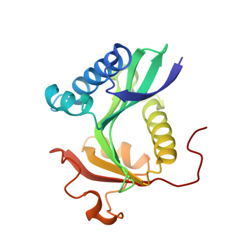
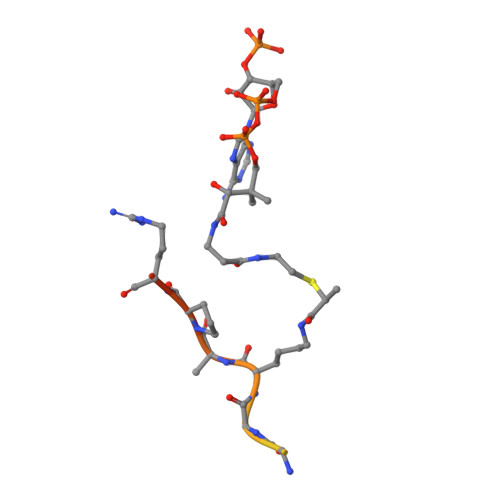
Structure of the GCN5 histone acetyltransferase bound to a bisubstrate inhibitor.

Poux, A.N.Cebrat, M.Kim, C.M.Cole, P.A.Marmorstein, R.

(2002) Proc Natl Acad Sci U S A 99: 14065-14070

  • DOI: https://doi.org/10.1073/pnas.222373899
  • Primary Citation of Related Structures:  
    1M1D

  • PubMed Abstract: 

    Histone acetyltransferases (HATs) use acetyl CoA to acetylate target lysine residues within histones and other transcription factors, such as the p53 tumor suppressor, to promote gene activation. HAT enzymes fall into subfamilies with divergence in sequence and substrate preference. Several HAT proteins have been implicated in human cancer. We have previously reported on the preparation of peptide-CoA conjugate inhibitors with distinct specificities for the p300/CBP [cAMP response element binding protein (CREB)-binding protein] or GCN5 HAT subfamilies. Here we report on the crystal structure of the GCN5 HAT bound to a peptide-CoA conjugate containing CoA covalently attached through an isopropionyl linker to Lys-14 of a 20-aa N-terminal fragment of histone H3. Surprisingly, the structure reveals that the H3 portion of the inhibitor is bound outside of the binding site for the histone substrate and that only five of the 20 aa residues of the inhibitor are ordered. Rearrangements within the C-terminal region of the GCN5 protein appear to mediate this peptide displacement. Mutational and enzymatic data support the hypothesis that the observed structure corresponds to a late catalytic intermediate. The structure also provides a structural scaffold for the design of HAT-specific inhibitors that may have therapeutic applications for the treatment of HAT-mediated cancers.


  • Organizational Affiliation

    The Wistar Institute, and Department of Chemistry, University of Pennsylvania, Philadelphia, PA 19104, USA.


Macromolecules
Find similar proteins by:  (by identity cutoff)  |  3D Structure
Entity ID: 1
MoleculeChains Sequence LengthOrganismDetailsImage
TGCN5 HISTONE ACETYL TRANSFERASE
A, C
163Tetrahymena thermophilaMutation(s): 0 
EC: 2.3.1
UniProt
Find proteins for Q27198 (Tetrahymena thermophila)
Explore Q27198 
Go to UniProtKB:  Q27198
Entity Groups  
Sequence Clusters30% Identity50% Identity70% Identity90% Identity95% Identity100% Identity
UniProt GroupQ27198
Sequence Annotations
Expand
  • Reference Sequence

Find similar proteins by:  Sequence   |   3D Structure  

Entity ID: 2
MoleculeChains Sequence LengthOrganismDetailsImage
HISTONE H3
B, D
20N/AMutation(s): 1 
UniProt
Find proteins for P61830 (Saccharomyces cerevisiae (strain ATCC 204508 / S288c))
Explore P61830 
Go to UniProtKB:  P61830
Entity Groups  
Sequence Clusters30% Identity50% Identity70% Identity90% Identity95% Identity100% Identity
UniProt GroupP61830
Sequence Annotations
Expand
  • Reference Sequence
Small Molecules
Modified Residues  1 Unique
IDChains TypeFormula2D DiagramParent
LYX
Query on LYX
B, D
L-PEPTIDE LINKINGC30 H52 N9 O19 P3 SLYS
Experimental Data & Validation

Experimental Data

  • Method: X-RAY DIFFRACTION
  • Resolution: 2.20 Å
  • R-Value Free: 0.265 
  • R-Value Work: 0.213 
  • R-Value Observed: 0.260 
  • Space Group: P 21 21 21
Unit Cell:
Length ( Å )Angle ( ˚ )
a = 67.42α = 90
b = 67.83β = 90
c = 74.5γ = 90
Software Package:
Software NamePurpose
HKL-2000data collection
SCALEPACKdata scaling
AMoREphasing
CNSrefinement
HKL-2000data reduction

Structure Validation

View Full Validation Report


Entry History 

Deposition Data

Revision History  (Full details and data files)

  • Version 1.0: 2002-10-30
    Type: Initial release
  • Version 1.1: 2008-04-28
    Changes: Version format compliance
  • Version 1.2: 2011-07-13
    Changes: Version format compliance