1JBQ

STRUCTURE OF HUMAN CYSTATHIONINE BETA-SYNTHASE: A UNIQUE PYRIDOXAL 5'-PHOSPHATE DEPENDENT HEMEPROTEIN


Experimental Data Snapshot

  • Method: X-RAY DIFFRACTION
  • Resolution: 2.60 Å
  • R-Value Free: 0.296 
  • R-Value Work: 0.257 
  • R-Value Observed: 0.257 

wwPDB Validation   3D Report Full Report


Ligand Structure Quality Assessment 


This is version 1.3 of the entry. See complete history


Literature

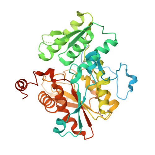
Structure of human cystathionine beta-synthase: a unique pyridoxal 5'-phosphate-dependent heme protein.

Meier, M.Janosik, M.Kery, V.Kraus, J.P.Burkhard, P.

(2001) EMBO J 20: 3910-3916

  • DOI: https://doi.org/10.1093/emboj/20.15.3910
  • Primary Citation of Related Structures:  
    1JBQ

  • PubMed Abstract: 

    Cystathionine beta-synthase (CBS) is a unique heme- containing enzyme that catalyzes a pyridoxal 5'-phosphate (PLP)-dependent condensation of serine and homocysteine to give cystathionine. Deficiency of CBS leads to homocystinuria, an inherited disease of sulfur metabolism characterized by increased levels of the toxic metabolite homocysteine. Here we present the X-ray crystal structure of a truncated form of the enzyme. CBS shares the same fold with O-acetylserine sulfhydrylase but it contains an additional N-terminal heme binding site. This heme binding motif together with a spatially adjacent oxidoreductase active site motif could explain the regulation of its enzyme activity by redox changes.


  • Organizational Affiliation

    M.E.Müller Institute for Structural Biology, Biozentrum, University of Basel, Klingelbergstrasse 70, CH-4056 Basel, Switzerland.


Macromolecules
Find similar proteins by:  (by identity cutoff)  |  3D Structure
Entity ID: 1
MoleculeChains Sequence LengthOrganismDetailsImage
CYSTATHIONINE BETA-SYNTHASE
A, B, C, D, E
A, B, C, D, E, F
435Homo sapiensMutation(s): 0 
EC: 4.2.1.22
UniProt & NIH Common Fund Data Resources
Find proteins for P35520 (Homo sapiens)
Explore P35520 
Go to UniProtKB:  P35520
PHAROS:  P35520
GTEx:  ENSG00000160200 
Entity Groups  
Sequence Clusters30% Identity50% Identity70% Identity90% Identity95% Identity100% Identity
UniProt GroupP35520
Sequence Annotations
Expand
  • Reference Sequence
Small Molecules
Ligands 2 Unique
IDChains Name / Formula / InChI Key2D Diagram3D Interactions
HEM
Query on HEM

Download Ideal Coordinates CCD File 
G [auth A]
I [auth B]
K [auth C]
M [auth D]
O [auth E]
G [auth A],
I [auth B],
K [auth C],
M [auth D],
O [auth E],
Q [auth F]
PROTOPORPHYRIN IX CONTAINING FE
C34 H32 Fe N4 O4
KABFMIBPWCXCRK-RGGAHWMASA-L
PLP
Query on PLP

Download Ideal Coordinates CCD File 
H [auth A]
J [auth B]
L [auth C]
N [auth D]
P [auth E]
H [auth A],
J [auth B],
L [auth C],
N [auth D],
P [auth E],
R [auth F]
PYRIDOXAL-5'-PHOSPHATE
C8 H10 N O6 P
NGVDGCNFYWLIFO-UHFFFAOYSA-N
Experimental Data & Validation

Experimental Data

  • Method: X-RAY DIFFRACTION
  • Resolution: 2.60 Å
  • R-Value Free: 0.296 
  • R-Value Work: 0.257 
  • R-Value Observed: 0.257 
  • Space Group: P 31
Unit Cell:
Length ( Å )Angle ( ˚ )
a = 144.52α = 90
b = 144.52β = 90
c = 108.16γ = 120
Software Package:
Software NamePurpose
DENZOdata reduction
SCALEPACKdata scaling
AMoREphasing
CNSrefinement

Structure Validation

View Full Validation Report


Ligand Structure Quality Assessment 


Entry History 

Deposition Data

Revision History  (Full details and data files)

  • Version 1.0: 2001-08-12
    Type: Initial release
  • Version 1.1: 2008-04-27
    Changes: Version format compliance
  • Version 1.2: 2011-07-13
    Changes: Non-polymer description, Version format compliance
  • Version 1.3: 2023-08-16
    Changes: Data collection, Database references, Derived calculations, Refinement description