1J78

Crystallographic analysis of the human vitamin D binding protein


Experimental Data Snapshot

  • Method: X-RAY DIFFRACTION
  • Resolution: 2.31 Å
  • R-Value Free: 0.252 
  • R-Value Work: 0.220 

wwPDB Validation   3D Report Full Report


Ligand Structure Quality Assessment 


This is version 1.3 of the entry. See complete history


Literature

A structural basis for the unique binding features of the human vitamin D-binding protein.

Verboven, C.Rabijns, A.De Maeyer, M.Van Baelen, H.Bouillon, R.De Ranter, C.

(2002) Nat Struct Biol 9: 131-136

  • DOI: https://doi.org/10.1038/nsb754
  • Primary Citation of Related Structures:  
    1J78, 1J7E

  • PubMed Abstract: 

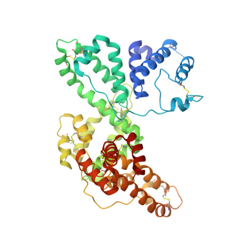
    The human serum vitamin D-binding protein (DBP) has many physiologically important functions, ranging from transporting vitamin D3 metabolites, binding and sequestering globular actin and binding fatty acids to functioning in the immune system. Here we report the 2.3 A crystal structure of DBP in complex with 25-hydroxyvitamin D3, a vitamin D3 metabolite, which reveals the vitamin D-binding site in the N-terminal part of domain I. To more explicitly explore this, we also studied the structure of DBP in complex with a vitamin D3 analog. Comparisons with the structure of human serum albumin, another family member, reveal a similar topology but also significant differences in overall, as well as local, folding. These observed structural differences explain the unique vitamin D3-binding property of DBP.


  • Organizational Affiliation

    Laboratorium voor Analytische Chemie en Medicinale Fysicochemie, Faculteit Farmaceutische Wetenschappen, Katholieke Universiteit Leuven, E. Van Evenstraat 4, B-3000 Leuven, Belgium.


Macromolecules
Find similar proteins by:  (by identity cutoff)  |  3D Structure
Entity ID: 1
MoleculeChains Sequence LengthOrganismDetailsImage
vitamin D binding protein
A, B
458Homo sapiensMutation(s): 1 
UniProt & NIH Common Fund Data Resources
Find proteins for P02774 (Homo sapiens)
Explore P02774 
Go to UniProtKB:  P02774
PHAROS:  P02774
GTEx:  ENSG00000145321 
Entity Groups  
Sequence Clusters30% Identity50% Identity70% Identity90% Identity95% Identity100% Identity
UniProt GroupP02774
Sequence Annotations
Expand
  • Reference Sequence
Experimental Data & Validation

Experimental Data

  • Method: X-RAY DIFFRACTION
  • Resolution: 2.31 Å
  • R-Value Free: 0.252 
  • R-Value Work: 0.220 
  • Space Group: P 43
Unit Cell:
Length ( Å )Angle ( ˚ )
a = 132.248α = 90
b = 132.248β = 90
c = 73.179γ = 90
Software Package:
Software NamePurpose
DENZOdata reduction
SCALEPACKdata scaling
SHARPphasing
CNSrefinement

Structure Validation

View Full Validation Report


Ligand Structure Quality Assessment 


Entry History 

Deposition Data

Revision History  (Full details and data files)

  • Version 1.0: 2002-02-06
    Type: Initial release
  • Version 1.1: 2008-04-27
    Changes: Version format compliance
  • Version 1.2: 2011-07-13
    Changes: Version format compliance
  • Version 1.3: 2018-01-31
    Changes: Experimental preparation