1IFA

THREE-DIMENSIONAL CRYSTAL STRUCTURE OF RECOMBINANT MURINE INTERFERON-BETA


Experimental Data Snapshot

  • Method: X-RAY DIFFRACTION
  • Resolution: 2.60 Å
  • R-Value Work: 0.205 
  • R-Value Observed: 0.205 

wwPDB Validation   3D Report Full Report


This is version 1.5 of the entry. See complete history


Literature

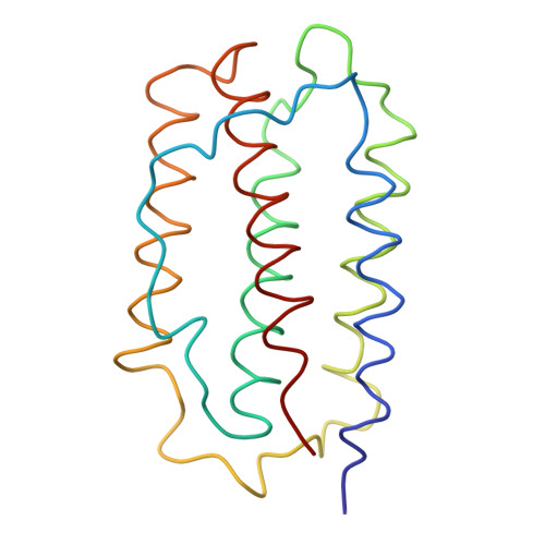
Three-dimensional crystal structure of recombinant murine interferon-beta.

Senda, T.Shimazu, T.Matsuda, S.Kawano, G.Shimizu, H.Nakamura, K.T.Mitsui, Y.

(1992) EMBO J 11: 3193-3201

  • DOI: https://doi.org/10.1002/j.1460-2075.1992.tb05396.x
  • Primary Citation of Related Structures:  
    1IFA

  • PubMed Abstract: 

    The crystal structure of recombinant murine interferon-beta (IFN-beta) has been solved by the multiple isomorphous replacement method and refined to an R-factor of 20.5% against 2.6 A X-ray diffraction data. The structure shows a variant of the alpha-helix bundle with a new chain-folding topology, which seems to represent a basic structural framework of all the IFN-alpha and IFN-beta molecules belonging to the type I family. Functionally important segments of the polypeptide chain, as implied through numerous gene manipulation studies carried out so far, are spatially clustered indicating the binding site(s) to the receptor(s). Comparison of the present structure with those of other alpha-helical cytokine proteins, including porcine growth hormone, interleukin 2 and interferon gamma, indicated either a topological similarity in chain folding or a similar spatial arrangement of the alpha-helices.


  • Organizational Affiliation

    Faculty of Engineering, Nagaoka University of Technology, Niigata, Japan.


Macromolecules
Find similar proteins by:  (by identity cutoff)  |  3D Structure
Entity ID: 1
MoleculeChains Sequence LengthOrganismDetailsImage
INTERFERON-BETA158Mus musculusMutation(s): 0 
UniProt
Find proteins for P01575 (Mus musculus)
Explore P01575 
Go to UniProtKB:  P01575
Entity Groups  
Sequence Clusters30% Identity50% Identity70% Identity90% Identity95% Identity100% Identity
UniProt GroupP01575
Sequence Annotations
Expand
  • Reference Sequence
Small Molecules
Ligands 1 Unique
IDChains Name / Formula / InChI Key2D Diagram3D Interactions
ASN
Query on ASN

Download Ideal Coordinates CCD File 
B [auth A]ASPARAGINE
C4 H8 N2 O3
DCXYFEDJOCDNAF-REOHCLBHSA-N
Experimental Data & Validation

Experimental Data

  • Method: X-RAY DIFFRACTION
  • Resolution: 2.60 Å
  • R-Value Work: 0.205 
  • R-Value Observed: 0.205 
  • Space Group: P 65
Unit Cell:
Length ( Å )Angle ( ˚ )
a = 71.4α = 90
b = 71.4β = 90
c = 79.6γ = 120
Software Package:
Software NamePurpose
X-PLORmodel building
PROLSQrefinement
X-PLORrefinement
X-PLORphasing

Structure Validation

View Full Validation Report


Entry History 

Deposition Data

Revision History  (Full details and data files)

  • Version 1.0: 1994-01-31
    Type: Initial release
  • Version 1.1: 2008-03-24
    Changes: Version format compliance
  • Version 1.2: 2011-07-13
    Changes: Version format compliance
  • Version 1.3: 2019-07-17
    Changes: Data collection, Other, Refinement description
  • Version 1.4: 2019-08-14
    Changes: Data collection, Refinement description
  • Version 1.5: 2024-02-07
    Changes: Data collection, Database references