1I7G

CRYSTAL STRUCTURE OF THE LIGAND BINDING DOMAIN FROM HUMAN PPAR-ALPHA IN COMPLEX WITH THE AGONIST AZ 242


Experimental Data Snapshot

  • Method: X-RAY DIFFRACTION
  • Resolution: 2.20 Å
  • R-Value Free: 0.271 
  • R-Value Work: 0.237 

wwPDB Validation   3D Report Full Report


This is version 1.3 of the entry. See complete history


Literature

Structure of the PPARalpha and -gamma ligand binding domain in complex with AZ 242; ligand selectivity and agonist activation in the PPAR family.

Cronet, P.Petersen, J.F.Folmer, R.Blomberg, N.Sjoblom, K.Karlsson, U.Lindstedt, E.L.Bamberg, K.

(2001) Structure 9: 699-706

  • DOI: https://doi.org/10.1016/s0969-2126(01)00634-7
  • Primary Citation of Related Structures:  
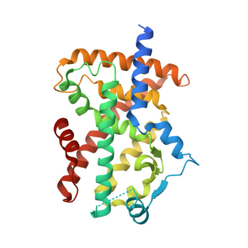
    1I7G, 1I7I

  • PubMed Abstract: 

    The peroxisome proliferator-activated receptors (PPAR) are ligand-activated transcription factors belonging to the nuclear receptor family. The roles of PPARalpha in fatty acid oxidation and PPARgamma in adipocyte differentiation and lipid storage have been characterized extensively. PPARs are activated by fatty acids and eicosanoids and are also targets for antidyslipidemic drugs, but the molecular interactions governing ligand selectivity for specific subtypes are unclear due to the lack of a PPARalpha ligand binding domain structure.


  • Organizational Affiliation

    Department of Molecular Biology, AstraZeneca R&D Mölndal, S-431 83, Mölndal, Sweden.


Macromolecules
Find similar proteins by:  (by identity cutoff)  |  3D Structure
Entity ID: 1
MoleculeChains Sequence LengthOrganismDetailsImage
PEROXISOME PROLIFERATOR ACTIVATED RECEPTOR ALPHA287Homo sapiensMutation(s): 0 
UniProt & NIH Common Fund Data Resources
Find proteins for Q07869 (Homo sapiens)
Explore Q07869 
Go to UniProtKB:  Q07869
PHAROS:  Q07869
GTEx:  ENSG00000186951 
Entity Groups  
Sequence Clusters30% Identity50% Identity70% Identity90% Identity95% Identity100% Identity
UniProt GroupQ07869
Sequence Annotations
Expand
  • Reference Sequence
Small Molecules
Ligands 3 Unique
IDChains Name / Formula / InChI Key2D Diagram3D Interactions
CPQ
Query on CPQ

Download Ideal Coordinates CCD File 
D [auth A]N,N-BIS(3-D-GLUCONAMIDOPROPYL)DEOXYCHOLAMIDE
C42 H75 N3 O15
OJSUWTDDXLCUFR-HGZMBBKESA-N
AZ2
Query on AZ2

Download Ideal Coordinates CCD File 
C [auth A](2S)-2-ETHOXY-3-[4-(2-{4-[(METHYLSULFONYL)OXY]PHENYL}ETHOXY)PHENYL]PROPANOIC ACID
C20 H24 O7 S
CXGTZJYQWSUFET-IBGZPJMESA-N
NA
Query on NA

Download Ideal Coordinates CCD File 
B [auth A]SODIUM ION
Na
FKNQFGJONOIPTF-UHFFFAOYSA-N
Binding Affinity Annotations 
IDSourceBinding Affinity
AZ2 BindingDB:  1I7G Ki: 1000 (nM) from 1 assay(s)
IC50: 3800 (nM) from 1 assay(s)
EC50: min: 37, max: 9798 (nM) from 10 assay(s)
PDBBind:  1I7G IC50: 1000 (nM) from 1 assay(s)
Binding MOAD:  1I7G IC50: 1000 (nM) from 1 assay(s)
Experimental Data & Validation

Experimental Data

  • Method: X-RAY DIFFRACTION
  • Resolution: 2.20 Å
  • R-Value Free: 0.271 
  • R-Value Work: 0.237 
  • Space Group: P 31 2 1
Unit Cell:
Length ( Å )Angle ( ˚ )
a = 76.968α = 90
b = 76.968β = 90
c = 100.592γ = 120
Software Package:
Software NamePurpose
EPMRphasing
CNXrefinement
MOSFLMdata reduction
CCP4data scaling

Structure Validation

View Full Validation Report


Entry History 

Deposition Data

Revision History  (Full details and data files)

  • Version 1.0: 2002-03-09
    Type: Initial release
  • Version 1.1: 2008-04-27
    Changes: Version format compliance
  • Version 1.2: 2011-07-13
    Changes: Version format compliance
  • Version 1.3: 2023-08-09
    Changes: Data collection, Database references, Derived calculations, Refinement description