1GH7

CRYSTAL STRUCTURE OF THE COMPLETE EXTRACELLULAR DOMAIN OF THE BETA-COMMON RECEPTOR OF IL-3, IL-5, AND GM-CSF


Experimental Data Snapshot

  • Method: X-RAY DIFFRACTION
  • Resolution: 3.00 Å
  • R-Value Free: 0.307 
  • R-Value Work: 0.267 

wwPDB Validation   3D Report Full Report


This is version 2.2 of the entry. See complete history


Literature

Structure of the complete extracellular domain of the common beta subunit of the human GM-CSF, IL-3, and IL-5 receptors reveals a novel dimer configuration.

Carr, P.D.Gustin, S.E.Church, A.P.Murphy, J.M.Ford, S.C.Mann, D.A.Woltring, D.M.Walker, I.Ollis, D.L.Young, I.G.

(2001) Cell 104: 291-300

  • DOI: https://doi.org/10.1016/s0092-8674(01)00213-6
  • Primary Citation of Related Structures:  
    1GH7

  • PubMed Abstract: 

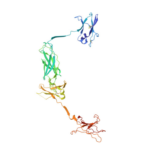
    The receptor systems for the hemopoietic cytokines GM-CSF, IL-3, and IL-5 consist of ligand-specific alpha receptor subunits that play an essential role in the activation of the shared betac subunit, the major signaling entity. Here, we report the structure of the complete betac extracellular domain. It has a structure unlike any class I cytokine receptor described thus far, forming a stable interlocking dimer in the absence of ligand in which the G strand of domain 1 hydrogen bonds into the corresponding beta sheet of domain 3 of the dimer-related molecule. The G strand of domain 3 similarly partners with the dimer-related domain 1. The structure provides new insights into receptor activation by the respective alpha receptor:ligand complexes.


  • Organizational Affiliation

    Research School of Chemistry, Australian National University, Acton, ACT 0200, Australia.


Macromolecules
Find similar proteins by:  (by identity cutoff)  |  3D Structure
Entity ID: 1
MoleculeChains Sequence LengthOrganismDetailsImage
CYTOKINE RECEPTOR COMMON BETA CHAIN
A, B
419Homo sapiensMutation(s): 1 
UniProt & NIH Common Fund Data Resources
Find proteins for P32927 (Homo sapiens)
Explore P32927 
Go to UniProtKB:  P32927
PHAROS:  P32927
GTEx:  ENSG00000100368 
Entity Groups  
Sequence Clusters30% Identity50% Identity70% Identity90% Identity95% Identity100% Identity
UniProt GroupP32927
Sequence Annotations
Expand
  • Reference Sequence
Oligosaccharides

Help

Entity ID: 2
MoleculeChains Length2D Diagram Glycosylation3D Interactions
beta-D-mannopyranose-(1-4)-2-acetamido-2-deoxy-beta-D-glucopyranose-(1-4)-2-acetamido-2-deoxy-beta-D-glucopyranose
C, E
3N-Glycosylation
Glycosylation Resources
GlyTouCan:  G15407YE
GlyCosmos:  G15407YE
GlyGen:  G15407YE
Entity ID: 3
MoleculeChains Length2D Diagram Glycosylation3D Interactions
2-acetamido-2-deoxy-beta-D-glucopyranose-(1-4)-[alpha-L-fucopyranose-(1-6)]2-acetamido-2-deoxy-beta-D-glucopyranose
D, F
3N-Glycosylation
Glycosylation Resources
GlyTouCan:  G21290RB
GlyCosmos:  G21290RB
GlyGen:  G21290RB
Experimental Data & Validation

Experimental Data

  • Method: X-RAY DIFFRACTION
  • Resolution: 3.00 Å
  • R-Value Free: 0.307 
  • R-Value Work: 0.267 
  • Space Group: H 3
Unit Cell:
Length ( Å )Angle ( ˚ )
a = 185.7α = 90
b = 185.7β = 90
c = 103.3γ = 120
Software Package:
Software NamePurpose
DENZOdata reduction
SCALEPACKdata scaling
CCP4model building
CNSrefinement
CCP4phasing

Structure Validation

View Full Validation Report


Entry History 

Deposition Data

Revision History  (Full details and data files)

  • Version 1.0: 2001-11-28
    Type: Initial release
  • Version 1.1: 2008-04-26
    Changes: Version format compliance
  • Version 1.2: 2011-07-13
    Changes: Non-polymer description, Version format compliance
  • Version 2.0: 2020-07-29
    Type: Remediation
    Reason: Carbohydrate remediation
    Changes: Advisory, Atomic model, Data collection, Database references, Derived calculations, Structure summary
  • Version 2.1: 2021-10-27
    Changes: Database references, Derived calculations, Structure summary
  • Version 2.2: 2023-12-27
    Changes: Data collection