1F4J

STRUCTURE OF TETRAGONAL CRYSTALS OF HUMAN ERYTHROCYTE CATALASE


Experimental Data Snapshot

  • Method: X-RAY DIFFRACTION
  • Resolution: 2.40 Å
  • R-Value Free: 0.244 
  • R-Value Work: 0.196 
  • R-Value Observed: 0.200 

wwPDB Validation   3D Report Full Report


Ligand Structure Quality Assessment 


This is version 1.4 of the entry. See complete history


Literature

Structure of tetragonal crystals of human erythrocyte catalase.

Safo, M.K.Musayev, F.N.Wu, S.H.Abraham, D.J.Ko, T.P.

(2001) Acta Crystallogr D Biol Crystallogr 57: 1-7

  • DOI: https://doi.org/10.1107/s0907444900013767
  • Primary Citation of Related Structures:  
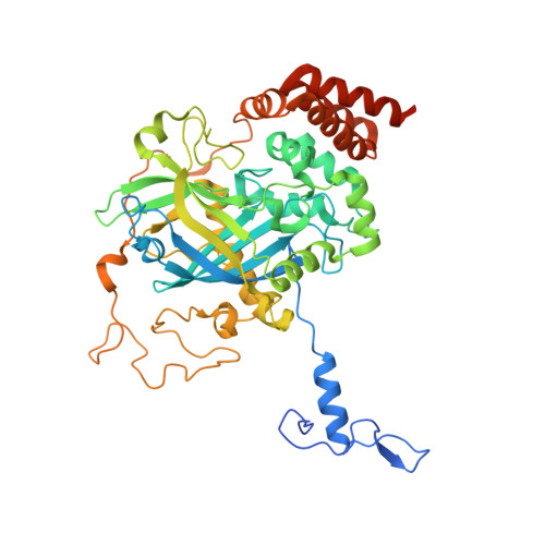
    1F4J

  • PubMed Abstract: 

    The structure of catalase from human erythrocytes (HEC) was determined in tetragonal crystals of space group I4(1) by molecular-replacement methods, using the orthorhombic crystal structure as a search model. It was then refined in a unit cell of dimensions a = b = 203.6 and c = 144.6 A, yielding R and R(free) of 0.196 and 0.244, respectively, for all data at 2.4 A resolution. A major difference of the HEC structure in the tetragonal crystal compared with the orthorhombic structure was the omission of a 20-residue N-terminal segment corresponding to the first exon of the human catalase gene. The overall structures were otherwise identical in both crystal forms. The NADPH-binding sites were empty in all four subunits and bound water molecules were observed at the active sites. The structure of the C-terminal segment, which corresponds to the last exon, remained undetermined. The tetragonal crystals showed a pseudo-4(1)22 symmetry in molecular packing. Two similar types of lattice contact interfaces between the HEC tetramers were observed; they were related by the pseudo-dyad axes.


  • Organizational Affiliation

    Institute for Structural Biology and Drug Discovery, Virginia Commonwealth University, Richmond, VA 23219, USA.


Macromolecules
Find similar proteins by:  (by identity cutoff)  |  3D Structure
Entity ID: 1
MoleculeChains Sequence LengthOrganismDetailsImage
CATALASE
A, B, C, D
527Homo sapiensMutation(s): 0 
EC: 1.11.1.6
UniProt & NIH Common Fund Data Resources
Find proteins for P04040 (Homo sapiens)
Explore P04040 
Go to UniProtKB:  P04040
PHAROS:  P04040
GTEx:  ENSG00000121691 
Entity Groups  
Sequence Clusters30% Identity50% Identity70% Identity90% Identity95% Identity100% Identity
UniProt GroupP04040
Sequence Annotations
Expand
  • Reference Sequence
Experimental Data & Validation

Experimental Data

  • Method: X-RAY DIFFRACTION
  • Resolution: 2.40 Å
  • R-Value Free: 0.244 
  • R-Value Work: 0.196 
  • R-Value Observed: 0.200 
  • Space Group: I 41
Unit Cell:
Length ( Å )Angle ( ˚ )
a = 203.6α = 90
b = 203.6β = 90
c = 144.6γ = 90
Software Package:
Software NamePurpose
AMoREphasing
CNSrefinement
bioteXdata reduction
bioteXdata scaling

Structure Validation

View Full Validation Report


Ligand Structure Quality Assessment 


Entry History 

Deposition Data

Revision History  (Full details and data files)

  • Version 1.0: 2000-06-21
    Type: Initial release
  • Version 1.1: 2008-04-27
    Changes: Version format compliance
  • Version 1.2: 2011-07-13
    Changes: Version format compliance
  • Version 1.3: 2017-10-04
    Changes: Refinement description
  • Version 1.4: 2024-02-07
    Changes: Data collection, Database references, Derived calculations