1CVS

CRYSTAL STRUCTURE OF A DIMERIC FGF2-FGFR1 COMPLEX


Experimental Data Snapshot

  • Method: X-RAY DIFFRACTION
  • Resolution: 2.80 Å
  • R-Value Free: 0.281 
  • R-Value Work: 0.240 
  • R-Value Observed: 0.240 

wwPDB Validation   3D Report Full Report


This is version 1.3 of the entry. See complete history


Literature

Structural basis for FGF receptor dimerization and activation.

Plotnikov, A.N.Schlessinger, J.Hubbard, S.R.Mohammadi, M.

(1999) Cell 98: 641-650

  • DOI: https://doi.org/10.1016/s0092-8674(00)80051-3
  • Primary Citation of Related Structures:  
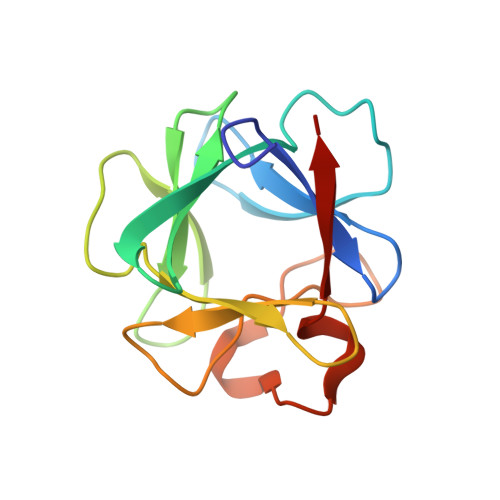
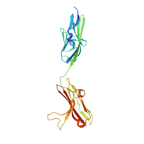
    1CVS

  • PubMed Abstract: 

    The crystal structure of FGF2 bound to a naturally occurring variant of FGF receptor 1 (FGFR1) consisting of immunoglobulin-like domains 2 (D2) and 3 (D3) has been determined at 2.8 A resolution. Two FGF2:FGFR1 complexes form a 2-fold symmetric dimer. Within each complex, FGF2 interacts extensively with D2 and D3 as well as with the linker between the two domains. The dimer is stabilized by interactions between FGF2 and D2 of the adjoining complex and by a direct interaction between D2 of each receptor. A positively charged canyon formed by a cluster of exposed basic residues likely represents the heparin-binding site. A general model for FGF- and heparin-induced FGFR dimerization is inferred from the crystal structure, unifying a wealth of biochemical data.


  • Organizational Affiliation

    Department of Pharmacology, New York University School of Medicine, New York 10016, USA.


Macromolecules
Find similar proteins by:  (by identity cutoff)  |  3D Structure
Entity ID: 1
MoleculeChains Sequence LengthOrganismDetailsImage
FIBROBLAST GROWTH FACTOR 2
A, B
132Homo sapiensMutation(s): 2 
UniProt & NIH Common Fund Data Resources
Find proteins for P09038 (Homo sapiens)
Explore P09038 
Go to UniProtKB:  P09038
PHAROS:  P09038
GTEx:  ENSG00000138685 
Entity Groups  
Sequence Clusters30% Identity50% Identity70% Identity90% Identity95% Identity100% Identity
UniProt GroupP09038
Sequence Annotations
Expand
  • Reference Sequence
Find similar proteins by:  (by identity cutoff)  |  3D Structure
Entity ID: 2
MoleculeChains Sequence LengthOrganismDetailsImage
FIBROBLAST GROWTH FACTOR RECEPTOR 1
C, D
225Homo sapiensMutation(s): 1 
UniProt & NIH Common Fund Data Resources
Find proteins for P11362 (Homo sapiens)
Explore P11362 
Go to UniProtKB:  P11362
PHAROS:  P11362
GTEx:  ENSG00000077782 
Entity Groups  
Sequence Clusters30% Identity50% Identity70% Identity90% Identity95% Identity100% Identity
UniProt GroupP11362
Sequence Annotations
Expand
  • Reference Sequence
Experimental Data & Validation

Experimental Data

  • Method: X-RAY DIFFRACTION
  • Resolution: 2.80 Å
  • R-Value Free: 0.281 
  • R-Value Work: 0.240 
  • R-Value Observed: 0.240 
  • Space Group: P 41 21 2
Unit Cell:
Length ( Å )Angle ( ˚ )
a = 98.45α = 90
b = 98.45β = 90
c = 197.03γ = 90
Software Package:
Software NamePurpose
SDMSdata collection
SCALEPACKdata scaling
AMoREphasing
CNSrefinement
SDMSdata reduction

Structure Validation

View Full Validation Report


Entry History 

Deposition Data

Revision History  (Full details and data files)

  • Version 1.0: 2000-01-28
    Type: Initial release
  • Version 1.1: 2008-04-27
    Changes: Version format compliance
  • Version 1.2: 2011-07-13
    Changes: Version format compliance
  • Version 1.3: 2024-04-03
    Changes: Data collection, Database references, Derived calculations, Refinement description