1BAK

SIGNAL TRANSDUCTION PLECKSTRIN HOMOLOGY DOMAIN OF G-PROTEIN COUPLED RECEPTOR KINASE 2 (BETA-ADRENERGIC RECEPTOR KINASE 1), C-TERMINAL EXTENDED, NMR, 20 STRUCTURES


Experimental Data Snapshot

  • Method: SOLUTION NMR
  • Conformers Calculated: 400 
  • Conformers Submitted: 20 
  • Selection Criteria: LEAST RESTRAINT VIOLATION 

wwPDB Validation   3D Report Full Report


This is version 1.3 of the entry. See complete history


Literature

The solution structure and dynamics of the pleckstrin homology domain of G protein-coupled receptor kinase 2 (beta-adrenergic receptor kinase 1). A binding partner of Gbetagamma subunits.

Fushman, D.Najmabadi-Haske, T.Cahill, S.Zheng, J.LeVine 3rd., H.Cowburn, D.

(1998) J Biol Chem 273: 2835-2843

  • DOI: https://doi.org/10.1074/jbc.273.5.2835
  • Primary Citation of Related Structures:  
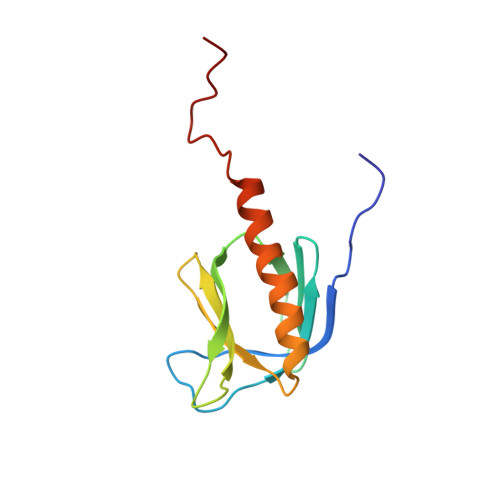
    1BAK

  • PubMed Abstract: 

    The solution structure of an extended pleckstrin homology (PH) domain from the beta-adrenergic receptor kinase is obtained by high resolution NMR. The structure establishes that the beta-adrenergic receptor kinase extended PH domain has the same fold and topology as other PH domains, and there are several unique features, most notably an extended C-terminal alpha-helix that behaves as a molten helix, and a surface charge polarity that is extensively modified by positive residues in the extended alpha-helix and the C terminus. These observations complement biochemical evidence that the C-terminal portion of this PH domain participates in protein-protein interactions with Gbetagamma subunits. This suggests that the C-terminal segment of the PH domain may function to mediate protein-protein interactions with the targets of PH domains.


  • Organizational Affiliation

    Laboratory of Physical Biochemistry, The Rockefeller University, New York, New York 10021-6399, USA.


Macromolecules
Find similar proteins by:  (by identity cutoff)  |  3D Structure
Entity ID: 1
MoleculeChains Sequence LengthOrganismDetailsImage
G-PROTEIN COUPLED RECEPTOR KINASE 2119Homo sapiensMutation(s): 0 
EC: 2.7.1.126
Membrane Entity: Yes 
UniProt & NIH Common Fund Data Resources
Find proteins for P25098 (Homo sapiens)
Explore P25098 
Go to UniProtKB:  P25098
PHAROS:  P25098
GTEx:  ENSG00000173020 
Entity Groups  
Sequence Clusters30% Identity50% Identity70% Identity90% Identity95% Identity100% Identity
UniProt GroupP25098
Sequence Annotations
Expand
  • Reference Sequence
Experimental Data & Validation

Experimental Data

  • Method: SOLUTION NMR
  • Conformers Calculated: 400 
  • Conformers Submitted: 20 
  • Selection Criteria: LEAST RESTRAINT VIOLATION 

Structure Validation

View Full Validation Report


Entry History 

Deposition Data

Revision History  (Full details and data files)

  • Version 1.0: 1998-02-25
    Type: Initial release
  • Version 1.1: 2008-03-24
    Changes: Version format compliance
  • Version 1.2: 2011-07-13
    Changes: Version format compliance
  • Version 1.3: 2022-02-16
    Changes: Data collection, Database references, Derived calculations, Other