5EX9

Structure of P450 StaF from glycopeptide antibiotic A47934 biosynthesis; glycerol cryo


Experimental Data Snapshot

  • Method: X-RAY DIFFRACTION
  • Resolution: 2.20 Å
  • R-Value Free: 0.220 
  • R-Value Work: 0.191 
  • R-Value Observed: 0.192 

wwPDB Validation   3D Report Full Report


Ligand Structure Quality Assessment 


This is version 1.2 of the entry. See complete history


Literature

Biochemical and structural characterisation of the second oxidative crosslinking step during the biosynthesis of the glycopeptide antibiotic A47934.

Ulrich, V.Brieke, C.Cryle, M.J.

(2016) Beilstein J Org Chem 12: 2849-2864

  • DOI: https://doi.org/10.3762/bjoc.12.284
  • Primary Citation of Related Structures:  
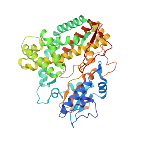
    5EX8, 5EX9

  • PubMed Abstract: 

    The chemical complexity and biological activity of the glycopeptide antibiotics (GPAs) stems from their unique crosslinked structure, which is generated by the actions of cytochrome P450 (Oxy) enzymes that affect the crosslinking of aromatic side chains of amino acid residues contained within the GPA heptapeptide precursor. Given the crucial role peptide cyclisation plays in GPA activity, the characterisation of this process is of great importance in understanding the biosynthesis of these important antibiotics. Here, we report the cyclisation activity and crystal structure of StaF, the D- O -E ring forming Oxy enzyme from A47934 biosynthesis. Our results show that the specificity of StaF is reduced when compared to Oxy enzymes catalysing C- O -D ring formation and that this activity relies on interactions with the non-ribosomal peptide synthetase via the X-domain. Despite the interaction of StaF with the A47934 X-domain being weaker than for the preceding Oxy enzyme StaH, StaF retains higher levels of in vitro activity: we postulate that this is due to the ability of the StaF/X-domain complex to allow substrate reorganisation after initial complex formation has occurred. These results highlight the importance of testing different peptide/protein carrier constructs for in vitro GPA cyclisation assays and show that different Oxy homologues can display significantly different catalytic propensities despite their overall similarities.


  • Organizational Affiliation

    Department of Biomolecular Mechanisms, Max Planck Institute for Medical Research, Jahnstrasse 29, 69120 Heidelberg, Germany.


Macromolecules
Find similar proteins by:  (by identity cutoff)  |  3D Structure
Entity ID: 1
MoleculeChains Sequence LengthOrganismDetailsImage
Cytochrome P450424Streptomyces toyocaensisMutation(s): 0 
Gene Names: BU52_01275
UniProt
Find proteins for Q8KLL7 (Streptomyces toyocaensis)
Explore Q8KLL7 
Go to UniProtKB:  Q8KLL7
Entity Groups  
Sequence Clusters30% Identity50% Identity70% Identity90% Identity95% Identity100% Identity
UniProt GroupQ8KLL7
Sequence Annotations
Expand
  • Reference Sequence
Small Molecules
Ligands 2 Unique
IDChains Name / Formula / InChI Key2D Diagram3D Interactions
HEM
Query on HEM

Download Ideal Coordinates CCD File 
B [auth A]PROTOPORPHYRIN IX CONTAINING FE
C34 H32 Fe N4 O4
KABFMIBPWCXCRK-RGGAHWMASA-L
GOL
Query on GOL

Download Ideal Coordinates CCD File 
C [auth A]
D [auth A]
E [auth A]
F [auth A]
G [auth A]
C [auth A],
D [auth A],
E [auth A],
F [auth A],
G [auth A],
H [auth A],
I [auth A],
J [auth A]
GLYCEROL
C3 H8 O3
PEDCQBHIVMGVHV-UHFFFAOYSA-N
Experimental Data & Validation

Experimental Data

  • Method: X-RAY DIFFRACTION
  • Resolution: 2.20 Å
  • R-Value Free: 0.220 
  • R-Value Work: 0.191 
  • R-Value Observed: 0.192 
  • Space Group: P 31 2 1
Unit Cell:
Length ( Å )Angle ( ˚ )
a = 109.7α = 90
b = 109.7β = 90
c = 93.9γ = 120
Software Package:
Software NamePurpose
REFMACrefinement
XDSdata reduction
Cootmodel building
PHASERphasing
XSCALEdata scaling

Structure Validation

View Full Validation Report


Ligand Structure Quality Assessment 


Entry History & Funding Information

Deposition Data


Funding OrganizationLocationGrant Number
German Research FoundationGermanyCR 392/1-1

Revision History  (Full details and data files)

  • Version 1.0: 2017-01-11
    Type: Initial release
  • Version 1.1: 2017-03-01
    Changes: Database references
  • Version 1.2: 2024-01-10
    Changes: Author supporting evidence, Data collection, Database references, Refinement description