3V4E

Crystal Structure of the galactoside O-acetyltransferase in complex with CoA


Experimental Data Snapshot

  • Method: X-RAY DIFFRACTION
  • Resolution: 1.95 Å
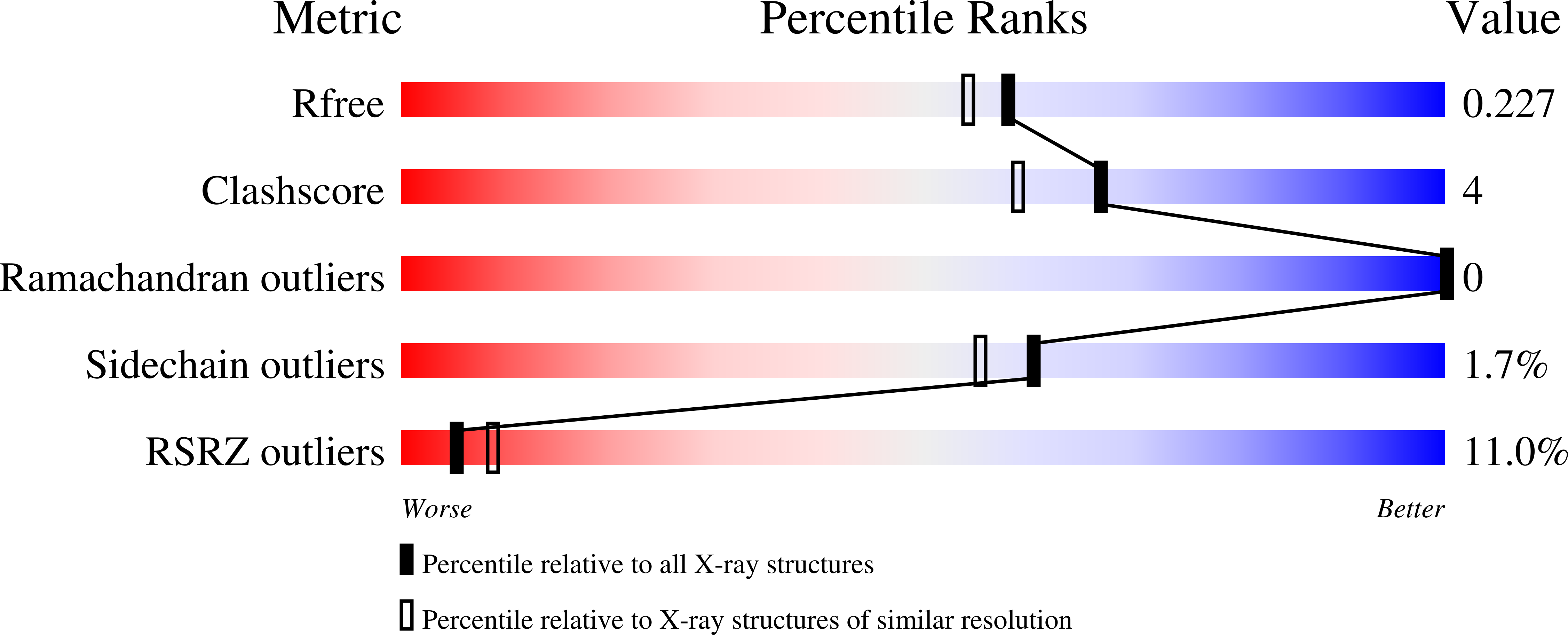
  • R-Value Free: 0.222 
  • R-Value Work: 0.180 
  • R-Value Observed: 0.183 

wwPDB Validation   3D Report Full Report


Ligand Structure Quality Assessment 


This is version 1.4 of the entry. See complete history


Literature

Biophysical analysis of the putative acetyltransferase SACOL2570 from methicillin-resistant Staphylococcus aureus.

Luo, H.B.Knapik, A.A.Petkowski, J.J.Demas, M.Shumilin, I.A.Zheng, H.Chruszcz, M.Minor, W.

(2013) J Struct Funct Genomics 14: 97-108

  • DOI: https://doi.org/10.1007/s10969-013-9158-6
  • Primary Citation of Related Structures:  
    3FTT, 3V4E

  • PubMed Abstract: 

    Methicillin-resistant Staphylococcus aureus (MRSA) is a major cause of a myriad of insidious and intractable infections in humans, especially in patients with compromised immune systems and children. Here, we report the apo- and CoA-bound crystal structures of a member of the galactoside acetyltransferase superfamily from methicillin-resistant S. aureus SACOL2570 which was recently shown to be down regulated in S. aureus grown in the presence of fusidic acid, an antibiotic used to treat MRSA infections. SACOL2570 forms a homotrimer in solution, as confirmed by small-angle X-ray scattering and dynamic light scattering. The protein subunit consists of an N-terminal alpha-helical domain connected to a C-terminal LβH domain. CoA binds in the active site formed by the residues from adjacent LβH domains. After determination of CoA-bound structure, molecular dynamics simulations were performed to model the binding of AcCoA. Binding of both AcCoA and CoA to SACOL2570 was verified by isothermal titration calorimetry. SACOL2570 most likely acts as an acetyltransferase, using AcCoA as an acetyl group donor and an as-yet-undetermined chemical moiety as an acceptor. SACOL2570 was recently used as a scaffold for mutations that lead the generation of cage-like assemblies, and has the potential to be used for the generation of more complex nanostructures.


  • Organizational Affiliation

    Department of Molecular Physiology and Biological Physics, University of Virginia, 1340 Jefferson Park Avenue, Charlottesville, VA 22908, USA.


Macromolecules
Find similar proteins by:  (by identity cutoff)  |  3D Structure
Entity ID: 1
MoleculeChains Sequence LengthOrganismDetailsImage
Galactoside O-acetyltransferase
A, B, C
202Staphylococcus aureus subsp. aureus COLMutation(s): 0 
Gene Names: SACOL2570
EC: 2.3.1
UniProt
Find proteins for Q5HCZ5 (Staphylococcus aureus (strain COL))
Explore Q5HCZ5 
Go to UniProtKB:  Q5HCZ5
Entity Groups  
Sequence Clusters30% Identity50% Identity70% Identity90% Identity95% Identity100% Identity
UniProt GroupQ5HCZ5
Sequence Annotations
Expand
  • Reference Sequence
Experimental Data & Validation

Experimental Data

  • Method: X-RAY DIFFRACTION
  • Resolution: 1.95 Å
  • R-Value Free: 0.222 
  • R-Value Work: 0.180 
  • R-Value Observed: 0.183 
  • Space Group: P 1
  • Diffraction Data: https://doi.org/10.18430/m30p4b
Unit Cell:
Length ( Å )Angle ( ˚ )
a = 53.424α = 89.75
b = 53.461β = 89.78
c = 53.477γ = 89.94
Software Package:
Software NamePurpose
MD2data collection
HKL-3000phasing
MOLREPphasing
REFMACrefinement
Cootmodel building
HKL-2000data reduction
HKL-2000data scaling

Structure Validation

View Full Validation Report


Ligand Structure Quality Assessment 


Entry History 

Revision History  (Full details and data files)

  • Version 1.0: 2012-01-11
    Type: Initial release
  • Version 1.1: 2017-02-15
    Changes: Database references
  • Version 1.2: 2017-11-08
    Changes: Refinement description
  • Version 1.3: 2022-04-13
    Changes: Database references, Derived calculations, Structure summary
  • Version 1.4: 2023-09-13
    Changes: Data collection, Refinement description