3DI0

Crystal Structure of Dihydrodipicolinate synthase from Staphylococcus aureus


Experimental Data Snapshot

  • Method: X-RAY DIFFRACTION
  • Resolution: 2.38 Å
  • R-Value Free: 0.250 
  • R-Value Work: 0.203 
  • R-Value Observed: 0.205 

wwPDB Validation   3D Report Full Report


This is version 1.2 of the entry. See complete history


Literature

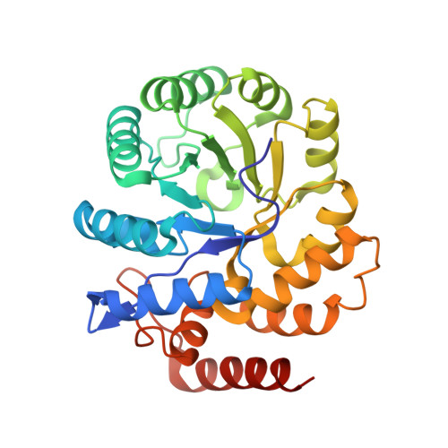
Structural and functional characterization of Staphylococcus aureus dihydrodipicolinate synthase

Girish, T.S.Sharma, E.Gopal, B.

(2008) FEBS Lett 582: 2923-2930

  • DOI: https://doi.org/10.1016/j.febslet.2008.07.035
  • Primary Citation of Related Structures:  
    3DI0, 3DI1

  • PubMed Abstract: 

    Lysine biosynthesis is crucial for cell-wall formation in bacteria. Enzymes involved in lysine biosynthesis are thus potential targets for anti-microbial therapeutics. Dihydrodipicolinate synthase (DHDPS) catalyzes the first step of this pathway. Unlike its homologues, Staphylococcus aureus DHDPS is a dimer both in solution and in the crystal and is not feedback inhibited by lysine. The crystal structure of S. aureus DHDPS in the free and substrate bound forms provides a structural rationale for its catalytic mechanism. The structure also reveals unique conformational features of the S. aureus enzyme that could be crucial for the design of specific non-competitive inhibitors.


  • Organizational Affiliation

    Molecular Biophysics Unit, Indian Institute of Science, Bangalore, India.


Macromolecules
Find similar proteins by:  (by identity cutoff)  |  3D Structure
Entity ID: 1
MoleculeChains Sequence LengthOrganismDetailsImage
Dihydrodipicolinate synthase
A, B
295Staphylococcus aureus subsp. aureus COLMutation(s): 0 
Gene Names: DHDPS
EC: 4.2.1.52
UniProt
Find proteins for Q5HG25 (Staphylococcus aureus (strain COL))
Explore Q5HG25 
Go to UniProtKB:  Q5HG25
Entity Groups  
Sequence Clusters30% Identity50% Identity70% Identity90% Identity95% Identity100% Identity
UniProt GroupQ5HG25
Sequence Annotations
Expand
  • Reference Sequence
Experimental Data & Validation

Experimental Data

  • Method: X-RAY DIFFRACTION
  • Resolution: 2.38 Å
  • R-Value Free: 0.250 
  • R-Value Work: 0.203 
  • R-Value Observed: 0.205 
  • Space Group: P 64 2 2
Unit Cell:
Length ( Å )Angle ( ˚ )
a = 118.686α = 90
b = 118.686β = 90
c = 179.156γ = 120
Software Package:
Software NamePurpose
REFMACrefinement
MAR345dtbdata collection
MOSFLMdata reduction
SCALAdata scaling
PHASERphasing

Structure Validation

View Full Validation Report


Entry History 

Deposition Data

Revision History  (Full details and data files)

  • Version 1.0: 2008-08-12
    Type: Initial release
  • Version 1.1: 2011-07-13
    Changes: Advisory, Source and taxonomy, Version format compliance
  • Version 1.2: 2023-11-01
    Changes: Data collection, Database references, Derived calculations, Refinement description