2BVE

Structure of the N-terminal of Sialoadhesin in complex with 2-Phenyl- Prop5Ac


Experimental Data Snapshot

  • Method: X-RAY DIFFRACTION
  • Resolution: 2.20 Å
  • R-Value Free: 0.270 
  • R-Value Work: 0.201 
  • R-Value Observed: 0.201 

wwPDB Validation   3D Report Full Report


This is version 1.4 of the entry. See complete history


Literature

Crystallographic and in Silico Analysis of the Sialoside-Binding Characteristics of the Siglec Sialoadhesin.

Zaccai, N.R.May, A.P.Robinson, R.C.Burtnick, L.D.Crocker, P.Brossmer, R.Kelm, S.Jones, E.Y.

(2007) J Mol Biol 365: 1469

  • DOI: https://doi.org/10.1016/j.jmb.2006.10.084
  • Primary Citation of Related Structures:  
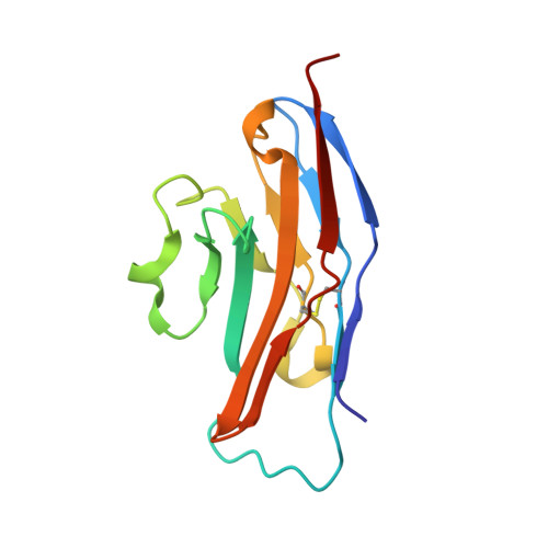
    2BVE

  • PubMed Abstract: 

    The Siglec family of receptors mediates cell-surface interactions through recognition of sialylated glycoconjugates. Previously reported structures of the N-terminal domain of the Siglec sialoadhesin (SnD1) in complex with various sialic acid analogs revealed the structural template for sialic acid binding. To characterize further the carbohydrate-binding properties, we have determined the crystal structures of SnD1 in the absence of ligand, and in complex with 2-benzyl-Neu5NPro and 2-benzyl-Neu5NAc. These structures reveal that SnD1 undergoes very few structural changes on ligand binding and detail how two novel classes of sialic acid analogs bind, one of which unexpectedly can induce Siglec dimerization. In conjunction with in silico analysis, this set of structures informs us about the design of putative ligands with enhanced binding affinities and specificities to different Siglecs, and provides data with which to test the effectiveness of different computational drug design protocols.


  • Organizational Affiliation

    CR-UK Receptor Structure Research Group, Division of Structural Biology, The Henry Wellcome Building for Genomic Medicine, Roosevelt Drive, Headington, Oxford OX3 7BN, UK. nathan.zaccai@bristol.ac.uk


Macromolecules
Find similar proteins by:  (by identity cutoff)  |  3D Structure
Entity ID: 1
MoleculeChains Sequence LengthOrganismDetailsImage
SIALOADHESIN
A, B
119Mus musculusMutation(s): 0 
UniProt & NIH Common Fund Data Resources
Find proteins for Q62230 (Mus musculus)
Explore Q62230 
Go to UniProtKB:  Q62230
IMPC:  MGI:99668
Entity Groups  
Sequence Clusters30% Identity50% Identity70% Identity90% Identity95% Identity100% Identity
UniProt GroupQ62230
Sequence Annotations
Expand
  • Reference Sequence
Small Molecules
Ligands 1 Unique
IDChains Name / Formula / InChI Key2D Diagram3D Interactions
PH5
Query on PH5

Download Ideal Coordinates CCD File 
C [auth A],
D [auth B]
benzyl 3,5-dideoxy-5-(propanoylamino)-D-glycero-alpha-D-galacto-non-2-ulopyranosidonic acid
C19 H27 N O9
XDURJXFZCXMSHL-YZKZVDITSA-N
Binding Affinity Annotations 
IDSourceBinding Affinity
PH5 PDBBind:  2BVE IC50: 2.15e+5 (nM) from 1 assay(s)
Binding MOAD:  2BVE IC50: 2.15e+5 (nM) from 1 assay(s)
Experimental Data & Validation

Experimental Data

  • Method: X-RAY DIFFRACTION
  • Resolution: 2.20 Å
  • R-Value Free: 0.270 
  • R-Value Work: 0.201 
  • R-Value Observed: 0.201 
  • Space Group: P 21 21 21
Unit Cell:
Length ( Å )Angle ( ˚ )
a = 31.974α = 90
b = 70.961β = 90
c = 102.65γ = 90
Software Package:
Software NamePurpose
CNSrefinement
DENZOdata reduction
SCALEPACKdata scaling
AMoREphasing

Structure Validation

View Full Validation Report


Entry History 

Deposition Data

Revision History  (Full details and data files)

  • Version 1.0: 2006-07-19
    Type: Initial release
  • Version 1.1: 2011-05-08
    Changes: Version format compliance
  • Version 1.2: 2011-07-13
    Changes: Version format compliance
  • Version 1.3: 2020-07-29
    Type: Remediation
    Reason: Carbohydrate remediation
    Changes: Derived calculations, Structure summary
  • Version 1.4: 2023-12-13
    Changes: Data collection, Database references, Refinement description, Structure summary