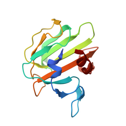
Crystal structures of active LytM.
Firczuk, M., Mucha, A., Bochtler, M.(2005) J Mol Biol 354: 578-590
- PubMed: 16269153
- DOI: https://doi.org/10.1016/j.jmb.2005.09.082
- Primary Citation of Related Structures:
2B0P, 2B13, 2B44 - PubMed Abstract:
Lysostaphin-type enzymes are metalloendopeptidases that are present in bacteriophages and in bacteria. They share the catalytic domain, but normally contain other domains as well. The well-characterized enzymes in this group are all specific for the pentaglycine crosslinks in the cell walls of some Gram-positive bacterial species. Lysostaphin-type enzymes are synthesized as secreted preproenzymes and require proteolytic activation for maturation. Although lysostaphin, the prototypical peptidase in the group, is widely used as a tool in biotechnology and developed as an antistaphylococcal agent, the detailed structure of this enzyme is unknown. So far, only one lysostaphin-type enzyme, the Staphylococcus aureus autolysin LytM, has been crystallized in its full-length, inactive form. Here, we describe the synthesis of a convenient reporter substrate, characterize the metal and pH-dependence of an active LytM fragment, and present its crystal structure in three crystal forms at different pH values that either support or do not support activity. In all structures, we find an extended, long and narrow groove that has the active site at its bottom and is delineated on the sides by the most flexible regions of the molecule. In two cases, the groove is partially filled by a loop of a neighbouring molecule in the crystal. As the loop contains three consecutive glycine residues, this crystal packing effect supports the interpretation that the groove is the substrate-binding cleft. To characterize the substrate-binding mode more closely, a phosphinate analogue of tetraglycine was synthesized. Although tetraglycine is a substrate of the active LytM fragment, the phosphinate analogue turned out to be a very poor inhibitor. Crystals that were grown in its presence contained an L+-tartrate molecule from the crystallization buffer and not the phosphinate in the active site.
Organizational Affiliation:
International Institute of Molecular and Cell Biology, ul. Trojdena 4, 02-109 Warsaw, Poland.