6OB1

Structure of WHB in complex with Ubiquitin Variant


Experimental Data Snapshot

  • Method: SOLUTION NMR
  • Conformers Calculated: 400 
  • Conformers Submitted: 20 
  • Selection Criteria: structures with the lowest energy 

wwPDB Validation   3D Report Full Report


This is version 1.2 of the entry. See complete history


Literature

Protein engineering of a ubiquitin-variant inhibitor of APC/C identifies a cryptic K48 ubiquitin chain binding site.

Watson, E.R.Grace, C.R.R.Zhang, W.Miller, D.J.Davidson, I.F.Prabu, J.R.Yu, S.Bolhuis, D.L.Kulko, E.T.Vollrath, R.Haselbach, D.Stark, H.Peters, J.M.Brown, N.G.Sidhu, S.S.Schulman, B.A.

(2019) Proc Natl Acad Sci U S A 116: 17280-17289

  • DOI: https://doi.org/10.1073/pnas.1902889116
  • Primary Citation of Related Structures:  
    6NXK, 6NXL, 6OB1

  • PubMed Abstract: 

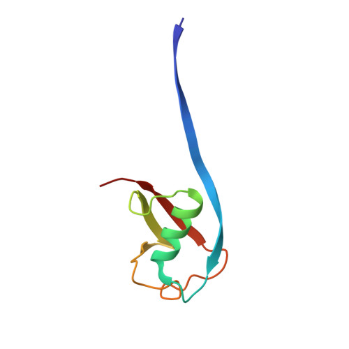
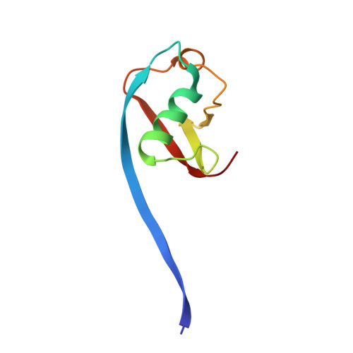
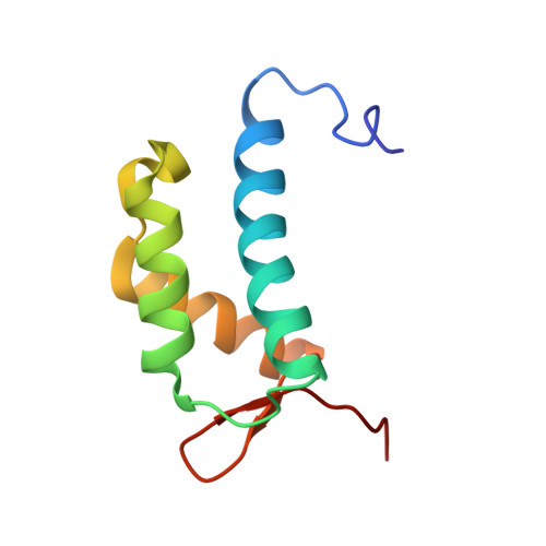
    Ubiquitin (Ub)-mediated proteolysis is a fundamental mechanism used by eukaryotic cells to maintain homeostasis and protein quality, and to control timing in biological processes. Two essential aspects of Ub regulation are conjugation through E1-E2-E3 enzymatic cascades and recognition by Ub-binding domains. An emerging theme in the Ub field is that these 2 properties are often amalgamated in conjugation enzymes. In addition to covalent thioester linkage to Ub's C terminus for Ub transfer reactions, conjugation enzymes often bind noncovalently and weakly to Ub at "exosites." However, identification of such sites is typically empirical and particularly challenging in large molecular machines. Here, studying the 1.2-MDa E3 ligase anaphase-promoting complex/cyclosome (APC/C), which controls cell division and many aspects of neurobiology, we discover a method for identifying unexpected Ub-binding sites. Using a panel of Ub variants (UbVs), we identify a protein-based inhibitor that blocks Ub ligation to APC/C substrates in vitro and ex vivo. Biochemistry, NMR, and cryo-electron microscopy (cryo-EM) structurally define the UbV interaction, explain its inhibitory activity through binding the surface on the APC2 subunit that recruits the E2 enzyme UBE2C, and ultimately reveal that this APC2 surface is also a Ub-binding exosite with preference for K48-linked chains. The results provide a tool for probing APC/C activity, have implications for the coordination of K48-linked Ub chain binding by APC/C with the multistep process of substrate polyubiquitylation, and demonstrate the power of UbV technology for identifying cryptic Ub-binding sites within large multiprotein complexes.


  • Organizational Affiliation

    Department of Structural Biology, St. Jude Children's Research Hospital, Memphis, TN 38105.


Macromolecules
Find similar proteins by:  (by identity cutoff)  |  3D Structure
Entity ID: 1
MoleculeChains Sequence LengthOrganismDetailsImage
Ubiquitin75Homo sapiensMutation(s): 0 
Entity Groups  
Sequence Clusters30% Identity50% Identity70% Identity90% Identity95% Identity100% Identity
Sequence Annotations
Expand
  • Reference Sequence
Find similar proteins by:  (by identity cutoff)  |  3D Structure
Entity ID: 2
MoleculeChains Sequence LengthOrganismDetailsImage
Ubiquitin75Homo sapiensMutation(s): 0 
Entity Groups  
Sequence Clusters30% Identity50% Identity70% Identity90% Identity95% Identity100% Identity
Sequence Annotations
Expand
  • Reference Sequence
Find similar proteins by:  (by identity cutoff)  |  3D Structure
Entity ID: 3
MoleculeChains Sequence LengthOrganismDetailsImage
Anaphase-promoting complex subunit 290Homo sapiensMutation(s): 0 
Gene Names: ANAPC2APC2KIAA1406
UniProt & NIH Common Fund Data Resources
Find proteins for Q9UJX6 (Homo sapiens)
Explore Q9UJX6 
Go to UniProtKB:  Q9UJX6
PHAROS:  Q9UJX6
GTEx:  ENSG00000176248 
Entity Groups  
Sequence Clusters30% Identity50% Identity70% Identity90% Identity95% Identity100% Identity
UniProt GroupQ9UJX6
Sequence Annotations
Expand
  • Reference Sequence
Experimental Data & Validation

Experimental Data

  • Method: SOLUTION NMR
  • Conformers Calculated: 400 
  • Conformers Submitted: 20 
  • Selection Criteria: structures with the lowest energy 

Structure Validation

View Full Validation Report


Entry History & Funding Information

Deposition Data


Funding OrganizationLocationGrant Number
National Institutes of Health/National Human Genome Research Institute (NIH/NHGRI)United StatesR35GM128855
National Institutes of Health/National Human Genome Research Institute (NIH/NHGRI)United StatesR37GM065930

Revision History  (Full details and data files)

  • Version 1.0: 2019-08-14
    Type: Initial release
  • Version 1.1: 2019-09-04
    Changes: Data collection, Database references
  • Version 1.2: 2019-12-18
    Changes: Author supporting evidence