6SK9

Nek2 bound to purine compound 51


Experimental Data Snapshot

  • Method: X-RAY DIFFRACTION
  • Resolution: 2.00 Å
  • R-Value Free: 0.220 
  • R-Value Work: 0.188 
  • R-Value Observed: 0.190 

wwPDB Validation   3D Report Full Report


Ligand Structure Quality Assessment 


This is version 1.0 of the entry. See complete history


Literature

Nek7 conformational flexibility and inhibitor binding probed through protein engineering of the R-spine.

Byrne, M.J.Nasir, N.Basmadjian, C.Bhatia, C.Cunnison, R.F.Carr, K.H.Mas-Droux, C.Yeoh, S.Cano, C.Bayliss, R.

(2020) Biochem J 477: 1525-1539

  • DOI: https://doi.org/10.1042/BCJ20200128
  • Primary Citation of Related Structures:  
    6S73, 6S75, 6S76, 6SK9

  • PubMed Abstract: 

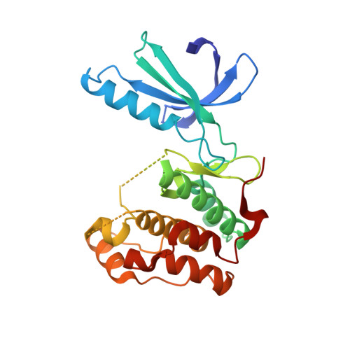
    Nek7 is a serine/threonine-protein kinase required for proper spindle formation and cytokinesis. Elevated Nek7 levels have been observed in several cancers, and inhibition of Nek7 might provide a route to the development of cancer therapeutics. To date, no selective and potent Nek7 inhibitors have been identified. Nek7 crystal structures exhibit an improperly formed regulatory-spine (R-spine), characteristic of an inactive kinase. We reasoned that the preference of Nek7 to crystallise in this inactive conformation might hinder attempts to capture Nek7 in complex with Type I inhibitors. Here, we have introduced aromatic residues into the R-spine of Nek7 with the aim to stabilise the active conformation of the kinase through R-spine stacking. The strong R-spine mutant Nek7SRS retained catalytic activity and was crystallised in complex with compound 51, an ATP-competitive inhibitor of Nek2 and Nek7. Subsequently, we obtained the same crystal form for wild-type Nek7WT in apo form and bound to compound 51. The R-spines of the three well-ordered Nek7WT molecules exhibit variable conformations while the R-spines of the Nek7SRS molecules all have the same, partially stacked configuration. Compound 51 bound to Nek2 and Nek7 in similar modes, but differences in the precise orientation of a substituent highlights features that could be exploited in designing inhibitors that are selective for particular Nek family members. Although the SRS mutations are not required to obtain a Nek7-inhibitor structure, we conclude that it is a useful strategy for restraining the conformation of a kinase in order to promote crystallogenesis.


  • Organizational Affiliation

    Astbury Centre for Structural Molecular Biology, School of Molecular and Cellular Biology, Faculty of Biological Sciences, University of Leeds, Leeds, U.K.


Macromolecules
Find similar proteins by:  (by identity cutoff)  |  3D Structure
Entity ID: 1
MoleculeChains Sequence LengthOrganismDetailsImage
Serine/threonine-protein kinase Nek2279Homo sapiensMutation(s): 0 
Gene Names: NEK2NEK2ANLK1
EC: 2.7.11.1
UniProt & NIH Common Fund Data Resources
Find proteins for P51955 (Homo sapiens)
Explore P51955 
Go to UniProtKB:  P51955
PHAROS:  P51955
GTEx:  ENSG00000117650 
Entity Groups  
Sequence Clusters30% Identity50% Identity70% Identity90% Identity95% Identity100% Identity
UniProt GroupP51955
Sequence Annotations
Expand
  • Reference Sequence
Small Molecules
Ligands 2 Unique
IDChains Name / Formula / InChI Key2D Diagram3D Interactions
F9N (Subject of Investigation/LOI)
Query on F9N

Download Ideal Coordinates CCD File 
B [auth A]3-[[6-(cyclohexylmethoxy)-7~{H}-purin-2-yl]amino]-~{N}-[3-(dimethylamino)propyl]benzenesulfonamide
C23 H33 N7 O3 S
PEEXIAQHZDNIBT-UHFFFAOYSA-N
GOL
Query on GOL

Download Ideal Coordinates CCD File 
C [auth A]GLYCEROL
C3 H8 O3
PEDCQBHIVMGVHV-UHFFFAOYSA-N
Binding Affinity Annotations 
IDSourceBinding Affinity
F9N Binding MOAD:  6SK9 IC50: 1420 (nM) from 1 assay(s)
Experimental Data & Validation

Experimental Data

  • Method: X-RAY DIFFRACTION
  • Resolution: 2.00 Å
  • R-Value Free: 0.220 
  • R-Value Work: 0.188 
  • R-Value Observed: 0.190 
  • Space Group: C 1 2 1
Unit Cell:
Length ( Å )Angle ( ˚ )
a = 100.02α = 90
b = 56.89β = 132.42
c = 77.94γ = 90
Software Package:
Software NamePurpose
PHENIXrefinement
MOSFLMdata reduction
SCALAdata scaling
PHASERphasing

Structure Validation

View Full Validation Report


Ligand Structure Quality Assessment 


Entry History & Funding Information

Deposition Data


Funding OrganizationLocationGrant Number
Cancer Research UKUnited KingdomC24461/ A13231
Medical Research Council (United Kingdom)United KingdomMR/L017032/1

Revision History  (Full details and data files)

  • Version 1.0: 2020-06-17
    Type: Initial release