6R2U

Zinc-alpha2-Glycoprotein with a Fluorescent Dansyl C 11 Fatty Acid


Experimental Data Snapshot

  • Method: X-RAY DIFFRACTION
  • Resolution: 2.49 Å
  • R-Value Free: 0.212 
  • R-Value Work: 0.170 
  • R-Value Observed: 0.171 

wwPDB Validation   3D Report Full Report


Ligand Structure Quality Assessment 


This is version 1.2 of the entry. See complete history


Literature

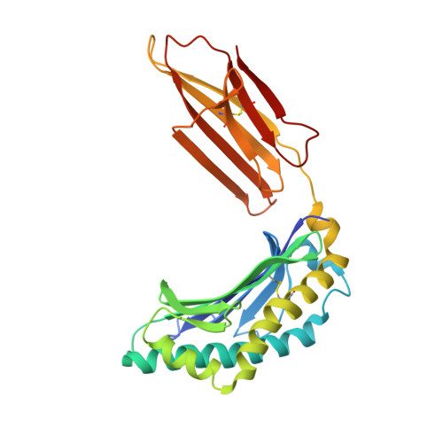
Crystal structure of zinc-alpha 2-glycoprotein in complex with a fatty acid reveals multiple different modes of protein-lipid binding.

Lau, A.M.Zahid, H.Gor, J.Perkins, S.J.Coker, A.R.McDermott, L.C.

(2019) Biochem J 476: 2815-2834

  • DOI: https://doi.org/10.1042/BCJ20190354
  • Primary Citation of Related Structures:  
    6R2U

  • PubMed Abstract: 

    Human zinc-α2-glycoprotein (ZAG) is a 42 kDa adipokine which regulates body fat mass and is associated with cachexia and obesity. ZAG belongs to the major histocompatibility complex class I protein family and binds long-chain polyunsaturated fatty acids in its groove formed from the α1 and α2 domains. To identify the molecular basis of its lipid-binding function, we determined the first crystal structure at 2.49 Å resolution for fatty acid-bound ZAG, where the ligand was the fluorescent 11-(dansylamino)undecanoic acid (DAUDA). The 192 kDa crystallographic asymmetric unit contained six ZAG and eight fatty acid molecules in unique conformations. Six fatty acid molecules were localised to the ZAG grooves, where their tails were bound in two distinct conformations. The carboxylate groups of three fatty acids projected out of the groove, while the fourth was hydrogen bonded with R73 inside the groove. Other ligand-residue contacts were primarily hydrophobic. A new fatty acid site was revealed for two further DAUDA molecules at the ZAG α3 domains. Following conformational changes from unbound ZAG, the α3 domains formed tetrameric β-barrel structures lined by fatty acid molecules that doubled the binding capacity of ZAG. Analytical ultracentrifugation revealed that ZAG in solution was a monomer in the absence of DAUDA, but formed small amounts of tetramers with DAUDA. By showing that ZAG binds fatty acids in different locations, we demonstrate an augmented mechanism for fatty acid binding in ZAG that is distinct from other known fatty acid binding proteins, and may be relevant to cachexia.


  • Organizational Affiliation

    Department of Structural and Molecular Biology, University College London, Darwin Building, Gower Street, London WC1E 6BT, U.K.


Macromolecules
Find similar proteins by:  (by identity cutoff)  |  3D Structure
Entity ID: 1
MoleculeChains Sequence LengthOrganismDetailsImage
Zinc-alpha-2-glycoprotein
A, B, C, D, E
A, B, C, D, E, F
279Homo sapiensMutation(s): 0 
Gene Names: AZGP1ZAGZNGP1
UniProt & NIH Common Fund Data Resources
Find proteins for P25311 (Homo sapiens)
Explore P25311 
Go to UniProtKB:  P25311
PHAROS:  P25311
GTEx:  ENSG00000160862 
Entity Groups  
Sequence Clusters30% Identity50% Identity70% Identity90% Identity95% Identity100% Identity
UniProt GroupP25311
Sequence Annotations
Expand
  • Reference Sequence
Small Molecules
Ligands 3 Unique
IDChains Name / Formula / InChI Key2D Diagram3D Interactions
11D (Subject of Investigation/LOI)
Query on 11D

Download Ideal Coordinates CCD File 
BA [auth F]
K [auth A]
L [auth B]
P [auth B]
Q [auth B]
BA [auth F],
K [auth A],
L [auth B],
P [auth B],
Q [auth B],
R [auth C],
V [auth D],
Z [auth E]
11-({[5-(dimethylamino)naphthalen-1-yl]sulfonyl}amino)undecanoic acid
C23 H34 N2 O4 S
CEPGVMDMVJGHFQ-UHFFFAOYSA-N
SO4
Query on SO4

Download Ideal Coordinates CCD File 
AA [auth E]
CA [auth F]
G [auth A]
H [auth A]
I [auth A]
AA [auth E],
CA [auth F],
G [auth A],
H [auth A],
I [auth A],
J [auth A],
N [auth B],
O [auth B],
S [auth C],
T [auth C],
U [auth C],
W [auth D],
X [auth D],
Y [auth D]
SULFATE ION
O4 S
QAOWNCQODCNURD-UHFFFAOYSA-L
AZI
Query on AZI

Download Ideal Coordinates CCD File 
M [auth B]AZIDE ION
N3
IVRMZWNICZWHMI-UHFFFAOYSA-N
Binding Affinity Annotations 
IDSourceBinding Affinity
11D Binding MOAD:  6R2U Kd: 510 (nM) from 1 assay(s)
Experimental Data & Validation

Experimental Data

  • Method: X-RAY DIFFRACTION
  • Resolution: 2.49 Å
  • R-Value Free: 0.212 
  • R-Value Work: 0.170 
  • R-Value Observed: 0.171 
  • Space Group: I 4
Unit Cell:
Length ( Å )Angle ( ˚ )
a = 167.015α = 90
b = 167.015β = 90
c = 204.948γ = 90
Software Package:
Software NamePurpose
REFMACrefinement
xia2data reduction
Aimlessdata scaling
PHASERphasing

Structure Validation

View Full Validation Report


Ligand Structure Quality Assessment 


Entry History 

Deposition Data

Revision History  (Full details and data files)

  • Version 1.0: 2019-09-18
    Type: Initial release
  • Version 1.1: 2019-11-06
    Changes: Data collection, Database references
  • Version 1.2: 2024-01-24
    Changes: Data collection, Database references, Refinement description