6PHY

SpAga D472N structure in complex alpha 1,3 galactobiose


Experimental Data Snapshot

  • Method: X-RAY DIFFRACTION
  • Resolution: 2.00 Å
  • R-Value Free: 0.216 
  • R-Value Work: 0.173 
  • R-Value Observed: 0.175 

wwPDB Validation   3D Report Full Report


This is version 2.1 of the entry. See complete history


Literature

Molecular analysis of an enigmaticStreptococcus pneumoniaevirulence factor: The raffinose-family oligosaccharide utilization system.

Hobbs, J.K.Meier, E.P.W.Pluvinage, B.Mey, M.A.Boraston, A.B.

(2019) J Biol Chem 294: 17197-17208

  • DOI: https://doi.org/10.1074/jbc.RA119.010280
  • Primary Citation of Related Structures:  
    6PHU, 6PHV, 6PHW, 6PHX, 6PHY, 6PI0, 6PQL, 6PRE, 6PRG

  • PubMed Abstract: 

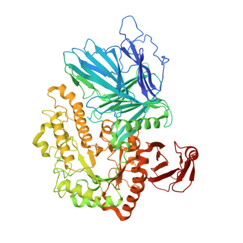
    Streptococcus pneumoniae is an opportunistic respiratory pathogen that can spread to other body sites, including the ears, brain, and blood. The ability of this bacterium to break down, import, and metabolize a wide range of glycans is key to its virulence. Intriguingly, S. pneumoniae can utilize several plant oligosaccharides for growth in vitro , including raffinose-family oligosaccharides (RFOs, which are α-(1→6)-galactosyl extensions of sucrose). An RFO utilization locus has been identified in the pneumococcal genome; however, none of the proteins encoded by this locus have been biochemically characterized. The enigmatic ability of S. pneumoniae to utilize RFOs has recently received attention because mutations in two of the RFO locus genes have been linked to the tissue tropism of clinical pneumococcal isolates. Here, we use functional studies combined with X-ray crystallography to show that although the pneumococcal RFO locus encodes for all the machinery required for uptake and degradation of RFOs, the individual pathway components are biochemically inefficient. We also demonstrate that the initiating enzyme in this pathway, the α-galactosidase Aga (a family 36 glycoside hydrolase), can cleave α-(1→3)-linked galactose units from a linear blood group antigen. We propose that the pneumococcal RFO pathway is an evolutionary relic that is not utilized in this streptococcal species and, as such, is under no selection pressure to maintain binding affinity and/or catalytic efficiency. We speculate that the apparent contribution of RFO utilization to pneumococcal tissue tropism may, in fact, be due to the essential role the ATPase RafK plays in the transport of other carbohydrates.


  • Organizational Affiliation

    Department of Biochemistry and Microbiology, University of Victoria, Victoria, British Columbia V8P 5C2, Canada.


Macromolecules
Find similar proteins by:  (by identity cutoff)  |  3D Structure
Entity ID: 1
MoleculeChains Sequence LengthOrganismDetailsImage
Alpha-galactosidase740Streptococcus pneumoniae TIGR4Mutation(s): 1 
Gene Names: agaSP_1898
EC: 3.2.1.22
UniProt
Find proteins for A0A0H2URQ6 (Streptococcus pneumoniae serotype 4 (strain ATCC BAA-334 / TIGR4))
Explore A0A0H2URQ6 
Go to UniProtKB:  A0A0H2URQ6
Entity Groups  
Sequence Clusters30% Identity50% Identity70% Identity90% Identity95% Identity100% Identity
UniProt GroupA0A0H2URQ6
Sequence Annotations
Expand
  • Reference Sequence
Oligosaccharides

Help

Entity ID: 2
MoleculeChains Length2D Diagram Glycosylation3D Interactions
alpha-D-galactopyranose-(1-3)-beta-D-galactopyranose
B
2N/A
Glycosylation Resources
GlyTouCan:  G24432YB
GlyCosmos:  G24432YB
GlyGen:  G24432YB
Entity ID: 3
MoleculeChains Length2D Diagram Glycosylation3D Interactions
alpha-D-galactopyranose-(1-3)-alpha-D-galactopyranose
C
2N/A
Glycosylation Resources
GlyTouCan:  G91563HD
GlyCosmos:  G91563HD
GlyGen:  G91563HD
Experimental Data & Validation

Experimental Data

  • Method: X-RAY DIFFRACTION
  • Resolution: 2.00 Å
  • R-Value Free: 0.216 
  • R-Value Work: 0.173 
  • R-Value Observed: 0.175 
  • Space Group: I 2 2 2
Unit Cell:
Length ( Å )Angle ( ˚ )
a = 90.958α = 90
b = 127.209β = 90
c = 152.136γ = 90
Software Package:
Software NamePurpose
HKL-2000data reduction
HKL-2000data scaling
PHASERphasing
REFMACrefinement
PDB_EXTRACTdata extraction

Structure Validation

View Full Validation Report


Entry History & Funding Information

Deposition Data


Funding OrganizationLocationGrant Number
Canadian Institutes of Health Research (CIHR)Canada--

Revision History  (Full details and data files)

  • Version 1.0: 2019-10-02
    Type: Initial release
  • Version 1.1: 2019-10-23
    Changes: Data collection, Database references
  • Version 1.2: 2019-11-27
    Changes: Database references
  • Version 1.3: 2020-01-08
    Changes: Author supporting evidence
  • Version 2.0: 2020-07-29
    Type: Remediation
    Reason: Carbohydrate remediation
    Changes: Atomic model, Author supporting evidence, Data collection, Derived calculations, Structure summary
  • Version 2.1: 2023-10-11
    Changes: Data collection, Database references, Refinement description, Structure summary