6O87

Crystal Structure of UDP-dependent glucosyltransferases (UGT) from Stevia rebaudiana in complex with UDP


Experimental Data Snapshot

  • Method: X-RAY DIFFRACTION
  • Resolution: 1.75 Å
  • R-Value Free: 0.194 
  • R-Value Work: 0.173 
  • R-Value Observed: 0.174 

wwPDB Validation   3D Report Full Report


Ligand Structure Quality Assessment 


This is version 1.3 of the entry. See complete history


Literature

Molecular basis for branched steviol glucoside biosynthesis.

Lee, S.G.Salomon, E.Yu, O.Jez, J.M.

(2019) Proc Natl Acad Sci U S A 116: 13131-13136

  • DOI: https://doi.org/10.1073/pnas.1902104116
  • Primary Citation of Related Structures:  
    6O86, 6O87, 6O88

  • PubMed Abstract: 

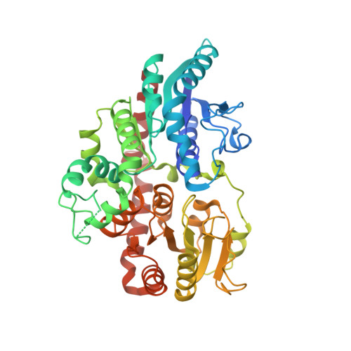
    Steviol glucosides, such as stevioside and rebaudioside A, are natural products roughly 200-fold sweeter than sugar and are used as natural, noncaloric sweeteners. Biosynthesis of rebaudioside A, and other related stevia glucosides, involves formation of the steviol diterpenoid followed by a series of glycosylations catalyzed by uridine diphosphate (UDP)-dependent glucosyltransferases. UGT76G1 from Stevia rebaudiana catalyzes the formation of the branched-chain glucoside that defines the stevia molecule and is critical for its high-intensity sweetness. Here, we report the 3D structure of the UDP-glucosyltransferase UGT76G1, including a complex of the protein with UDP and rebaudioside A bound in the active site. The X-ray crystal structure and biochemical analysis of site-directed mutants identifies a catalytic histidine and how the acceptor site of UGT76G1 achieves regioselectivity for branched-glucoside synthesis. The active site accommodates a two-glucosyl side chain and provides a site for addition of a third sugar molecule to the C3' position of the first C13 sugar group of stevioside. This structure provides insight on the glycosylation of other naturally occurring sweeteners, such as the mogrosides from monk fruit, and a possible template for engineering of steviol biosynthesis.


  • Organizational Affiliation

    Department of Biology, Washington University in St. Louis, St. Louis, MO 63130.


Macromolecules
Find similar proteins by:  (by identity cutoff)  |  3D Structure
Entity ID: 1
MoleculeChains Sequence LengthOrganismDetailsImage
UDP-glycosyltransferase 76G1458Stevia rebaudianaMutation(s): 0 
Gene Names: UGT76G1
EC: 2.4.1
UniProt
Find proteins for Q6VAB4 (Stevia rebaudiana)
Explore Q6VAB4 
Go to UniProtKB:  Q6VAB4
Entity Groups  
Sequence Clusters30% Identity50% Identity70% Identity90% Identity95% Identity100% Identity
UniProt GroupQ6VAB4
Sequence Annotations
Expand
  • Reference Sequence
Small Molecules
Ligands 1 Unique
IDChains Name / Formula / InChI Key2D Diagram3D Interactions
UDP (Subject of Investigation/LOI)
Query on UDP

Download Ideal Coordinates CCD File 
B [auth A]URIDINE-5'-DIPHOSPHATE
C9 H14 N2 O12 P2
XCCTYIAWTASOJW-XVFCMESISA-N
Experimental Data & Validation

Experimental Data

  • Method: X-RAY DIFFRACTION
  • Resolution: 1.75 Å
  • R-Value Free: 0.194 
  • R-Value Work: 0.173 
  • R-Value Observed: 0.174 
  • Space Group: P 31 2 1
Unit Cell:
Length ( Å )Angle ( ˚ )
a = 98.454α = 90
b = 98.454β = 90
c = 90.667γ = 120
Software Package:
Software NamePurpose
PHENIXrefinement
PHASERphasing
Cootmodel building
HKL-3000data collection

Structure Validation

View Full Validation Report


Ligand Structure Quality Assessment 


Entry History 

Deposition Data

Revision History  (Full details and data files)

  • Version 1.0: 2019-06-12
    Type: Initial release
  • Version 1.1: 2019-06-26
    Changes: Data collection, Database references
  • Version 1.2: 2019-07-10
    Changes: Data collection, Database references
  • Version 1.3: 2024-03-13
    Changes: Data collection, Database references