6N9T

Structure of a peptide-based photo-affinity cross-linker with Herceptin Fc


Experimental Data Snapshot

  • Method: X-RAY DIFFRACTION
  • Resolution: 2.58 Å
  • R-Value Free: 0.265 
  • R-Value Work: 0.229 
  • R-Value Observed: 0.231 

wwPDB Validation   3D Report Full Report


This is version 2.0 of the entry. See complete history


Literature

Development, Optimization, and Structural Characterization of an Efficient Peptide-Based Photoaffinity Cross-Linking Reaction for Generation of Homogeneous Conjugates from Wild-Type Antibodies.

Vance, N.Zacharias, N.Ultsch, M.Li, G.Fourie, A.Liu, P.LaFrance-Vanasse, J.Ernst, J.A.Sandoval, W.Kozak, K.R.Phillips, G.Wang, W.Sadowsky, J.

(2019) Bioconjug Chem 30: 148-160

  • DOI: https://doi.org/10.1021/acs.bioconjchem.8b00809
  • Primary Citation of Related Structures:  
    6N9T

  • PubMed Abstract: 

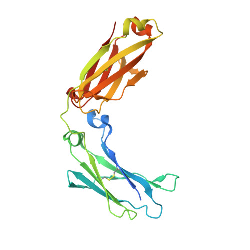
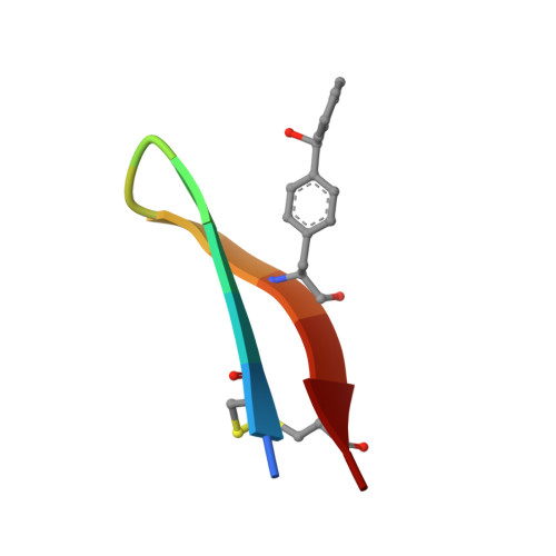
    Site-specific conjugation of small molecules to antibodies represents an attractive goal for the development of more homogeneous targeted therapies and diagnostics. Most site-specific conjugation strategies require modification or removal of antibody glycans or interchain disulfide bonds or engineering of an antibody mutant that bears a reactive handle. While such methods are effective, they complicate the process of preparing antibody conjugates and can negatively impact biological activity. Herein we report the development and detailed characterization of a robust photoaffinity cross-linking method for site-specific conjugation to fully glycosylated wild-type antibodies. The method employs a benzoylphenylalanine (Bpa) mutant of a previously described 13-residue peptide derived from phage display to bind tightly to the Fc domain; upon UV irradiation, the Bpa residue forms a diradical that reacts with the bound antibody. After the initial discovery of an effective Bpa mutant peptide and optimization of the reaction conditions to enable efficient conjugation without concomitant UV-induced photodamage of the antibody, we assessed the scope of the photoconjugation reaction across different human and nonhuman antibodies and antibody mutants. Next, the specific site of conjugation on a human antibody was characterized in detail by mass spectrometry experiments and at atomic resolution by X-ray crystallography. Finally, we adapted the photoconjugation method to attach a cytotoxic payload site-specifically to a wild-type antibody and showed that the resulting conjugate is both stable in plasma and as potent as a conventional antibody-drug conjugate in cells, portending well for future biological applications.


  • Organizational Affiliation

    Research & Early Development , Genentech, Inc. , 1 DNA Way , South San Francisco , California 94080 , United States.


Macromolecules
Find similar proteins by:  (by identity cutoff)  |  3D Structure
Entity ID: 1
MoleculeChains Sequence LengthOrganismDetailsImage
Immunoglobulin G1 FCA,
C [auth B]
224Homo sapiensMutation(s): 0 
UniProt
Find proteins for Q6MZV7 (Homo sapiens)
Explore Q6MZV7 
Go to UniProtKB:  Q6MZV7
Entity Groups  
Sequence Clusters30% Identity50% Identity70% Identity90% Identity95% Identity100% Identity
UniProt GroupQ6MZV7
Sequence Annotations
Expand
  • Reference Sequence

Find similar proteins by:  Sequence   |   3D Structure  

Entity ID: 2
MoleculeChains Sequence LengthOrganismDetailsImage
Photo-affinity peptideB [auth E],
D [auth F]
13synthetic constructMutation(s): 0 
Entity Groups  
Sequence Clusters30% Identity50% Identity70% Identity90% Identity95% Identity100% Identity
Sequence Annotations
Expand
  • Reference Sequence
Oligosaccharides

Help

Entity ID: 3
MoleculeChains Length2D Diagram Glycosylation3D Interactions
2-acetamido-2-deoxy-beta-D-glucopyranose-(1-2)-alpha-D-mannopyranose-(1-6)-[alpha-D-mannopyranose-(1-3)]beta-D-mannopyranose-(1-4)-2-acetamido-2-deoxy-beta-D-glucopyranose-(1-4)-[beta-L-fucopyranose-(1-6)]2-acetamido-2-deoxy-beta-D-glucopyranoseE [auth C]7N-Glycosylation
Glycosylation Resources
GlyTouCan:  G20449BJ
GlyCosmos:  G20449BJ
GlyGen:  G20449BJ
Entity ID: 4
MoleculeChains Length2D Diagram Glycosylation3D Interactions
2-acetamido-2-deoxy-beta-D-glucopyranose-(1-2)-alpha-D-mannopyranose-(1-6)-[alpha-D-mannopyranose-(1-3)]beta-D-mannopyranose-(1-4)-2-acetamido-2-deoxy-beta-D-glucopyranose-(1-4)-[alpha-L-fucopyranose-(1-6)]2-acetamido-2-deoxy-beta-D-glucopyranoseF [auth D]7N-Glycosylation
Glycosylation Resources
GlyTouCan:  G45889JQ
GlyCosmos:  G45889JQ
GlyGen:  G45889JQ
Small Molecules
Modified Residues  1 Unique
IDChains TypeFormula2D DiagramParent
PBF
Query on PBF
B [auth E],
D [auth F]
L-PEPTIDE LINKINGC16 H15 N O3PHE
Experimental Data & Validation

Experimental Data

  • Method: X-RAY DIFFRACTION
  • Resolution: 2.58 Å
  • R-Value Free: 0.265 
  • R-Value Work: 0.229 
  • R-Value Observed: 0.231 
  • Space Group: P 1 21 1
Unit Cell:
Length ( Å )Angle ( ˚ )
a = 66.112α = 90
b = 60.85β = 103.13
c = 68.167γ = 90
Software Package:
Software NamePurpose
PHENIXrefinement
XDSdata reduction
Aimlessdata scaling
PHASERphasing

Structure Validation

View Full Validation Report


Entry History 

Deposition Data

Revision History  (Full details and data files)

  • Version 1.0: 2019-01-16
    Type: Initial release
  • Version 1.1: 2019-05-01
    Changes: Data collection, Database references
  • Version 2.0: 2020-07-29
    Type: Remediation
    Reason: Carbohydrate remediation
    Changes: Advisory, Atomic model, Data collection, Derived calculations, Structure summary