The interaction of talin with the cell membrane is essential for integrin activation and focal adhesion formation.
Chinthalapudi, K., Rangarajan, E.S., Izard, T.(2018) Proc Natl Acad Sci U S A 115: 10339-10344
- PubMed: 30254158
- DOI: https://doi.org/10.1073/pnas.1806275115
- Primary Citation of Related Structures:
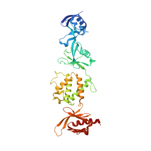
6MFS - PubMed Abstract:
Multicellular organisms have well-defined, tightly regulated mechanisms for cell adhesion. Heterodimeric αβ integrin receptors play central roles in this function and regulate processes for normal cell functions, including signaling, cell migration, and development, binding to the extracellular matrix, and senescence. They are involved in hemostasis and the immune response, participate in leukocyte function, and have biological implications in angiogenesis and cancer. Proper control of integrin activation for cellular communication with the external environment requires several physiological processes. Perturbation of these equilibria may lead to constitutive integrin activation that results in bleeding disorders. Furthermore, integrins play key roles in cancer progression and metastasis in which certain tumor types exhibit higher levels of various integrins. Thus, the integrin-associated signaling complex is important for cancer therapy development. During inside-out signaling, the cytoskeletal protein talin plays a key role in regulating integrin affinity whereby the talin head domain activates integrin by binding to the cytoplasmic tail of β-integrin and acidic membrane phospholipids. To understand the mechanism of integrin activation by talin, we determined the crystal structure of the talin head domain bound to the acidic phospholipid phosphatidylinositol 4,5-bisphosphate (PIP 2 ), allowing us to design a lipid-binding-deficient talin mutant. Our confocal microscopy with talin knockout cells suggests that the talin-cell membrane interaction seems essential for focal adhesion formation and stabilization. Basal integrin activation in Chinese hamster ovary cells suggests that the lipid-binding-deficient talin mutant inhibits integrin activation. Thus, membrane attachment of talin seems necessary for integrin activation and focal adhesion formation.
Organizational Affiliation:
Cell Adhesion Laboratory, Department of Integrative Structural and Computational Biology, The Scripps Research Institute, Jupiter, FL 33458.