6GWS

Crystal structure of human PCNA in complex with three p15 peptides


Experimental Data Snapshot

  • Method: X-RAY DIFFRACTION
  • Resolution: 2.90 Å
  • R-Value Free: 0.263 
  • R-Value Work: 0.200 
  • R-Value Observed: 0.203 

wwPDB Validation   3D Report Full Report


This is version 1.3 of the entry. See complete history


Literature

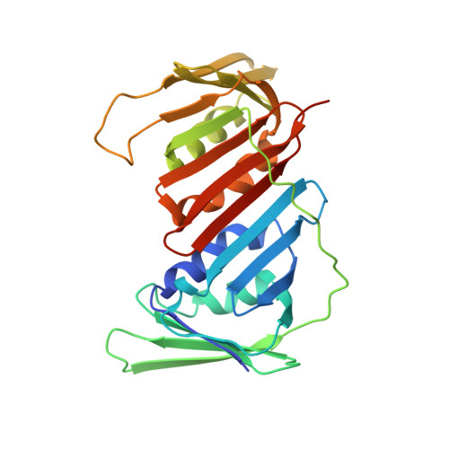
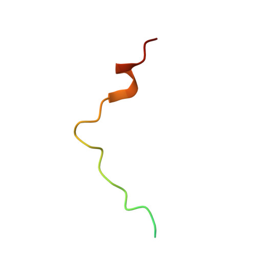
p15PAF binding to PCNA modulates the DNA sliding surface.

De March, M.Barrera-Vilarmau, S.Crespan, E.Mentegari, E.Merino, N.Gonzalez-Magana, A.Romano-Moreno, M.Maga, G.Crehuet, R.Onesti, S.Blanco, F.J.De Biasio, A.

(2018) Nucleic Acids Res 46: 9816-9828

  • DOI: https://doi.org/10.1093/nar/gky723
  • Primary Citation of Related Structures:  
    6EHT, 6GWS

  • PubMed Abstract: 

    p15PAF is an oncogenic intrinsically disordered protein that regulates DNA replication and lesion bypass by interacting with the human sliding clamp PCNA. In the absence of DNA, p15PAF traverses the PCNA ring via an extended PIP-box that contacts the sliding surface. Here, we probed the atomic-scale structure of p15PAF-PCNA-DNA ternary complexes. Crystallography and MD simulations show that, when p15PAF occupies two subunits of the PCNA homotrimer, DNA within the ring channel binds the unoccupied subunit. The structure of PCNA-bound p15PAF in the absence and presence of DNA is invariant, and solution NMR confirms that DNA does not displace p15PAF from the ring wall. Thus, p15PAF reduces the available sliding surfaces of PCNA, and may function as a belt that fastens the DNA to the clamp during synthesis by the replicative polymerase (pol δ). This constraint, however, may need to be released for efficient DNA lesion bypass by the translesion synthesis polymerase (pol η). Accordingly, our biochemical data show that p15PAF impairs primer synthesis by pol η-PCNA holoenzyme against both damaged and normal DNA templates. In light of our findings, we discuss the possible mechanistic roles of p15PAF in DNA replication and suppression of DNA lesion bypass.


  • Organizational Affiliation

    Structural Biology Laboratory, Elettra-Sincrotrone Trieste S.C.p.A., Trieste 34149, Italy.


Macromolecules
Find similar proteins by:  (by identity cutoff)  |  3D Structure
Entity ID: 1
MoleculeChains Sequence LengthOrganismDetailsImage
Proliferating cell nuclear antigen
A, B, C
264Homo sapiensMutation(s): 0 
Gene Names: PCNA
UniProt & NIH Common Fund Data Resources
Find proteins for P12004 (Homo sapiens)
Explore P12004 
Go to UniProtKB:  P12004
PHAROS:  P12004
GTEx:  ENSG00000132646 
Entity Groups  
Sequence Clusters30% Identity50% Identity70% Identity90% Identity95% Identity100% Identity
UniProt GroupP12004
Sequence Annotations
Expand
  • Reference Sequence
Find similar proteins by:  (by identity cutoff)  |  3D Structure
Entity ID: 2
MoleculeChains Sequence LengthOrganismDetailsImage
PCNA-associated factor
D, E, F
32Homo sapiensMutation(s): 0 
Gene Names: PCLAFKIAA0101NS5ATP9PAFL5
UniProt & NIH Common Fund Data Resources
Find proteins for Q15004 (Homo sapiens)
Explore Q15004 
Go to UniProtKB:  Q15004
PHAROS:  Q15004
GTEx:  ENSG00000166803 
Entity Groups  
Sequence Clusters30% Identity50% Identity70% Identity90% Identity95% Identity100% Identity
UniProt GroupQ15004
Sequence Annotations
Expand
  • Reference Sequence
Experimental Data & Validation

Experimental Data

  • Method: X-RAY DIFFRACTION
  • Resolution: 2.90 Å
  • R-Value Free: 0.263 
  • R-Value Work: 0.200 
  • R-Value Observed: 0.203 
  • Space Group: P 1 21 1
Unit Cell:
Length ( Å )Angle ( ˚ )
a = 79.24α = 90
b = 89.75β = 117.25
c = 85.13γ = 90
Software Package:
Software NamePurpose
XSCALEdata scaling
REFMACrefinement
PDB_EXTRACTdata extraction
XDSdata reduction
PHASERphasing

Structure Validation

View Full Validation Report


Entry History & Funding Information

Deposition Data


Funding OrganizationLocationGrant Number
Spanish Ministry of Economy and CompetitivenessSpainCTQ2017-83810-R

Revision History  (Full details and data files)

  • Version 1.0: 2018-08-22
    Type: Initial release
  • Version 1.1: 2018-09-05
    Changes: Data collection, Database references
  • Version 1.2: 2018-10-24
    Changes: Data collection, Database references
  • Version 1.3: 2024-01-17
    Changes: Data collection, Database references, Refinement description