Improved protein-crystal identification by using 2,2,2-trichloroethanol as a fluorescence enhancer.
Pichlo, C., Toelzer, C., Chojnacki, K., Ocal, S., Uthoff, M., Ruegenberg, S., Hermanns, T., Schacherl, M., Denzel, M.S., Hofmann, K., Niefind, K., Baumann, U.(2018) Acta Crystallogr F Struct Biol Commun 74: 307-314
- PubMed: 29717999
- DOI: https://doi.org/10.1107/S2053230X18005253
- Primary Citation of Related Structures:
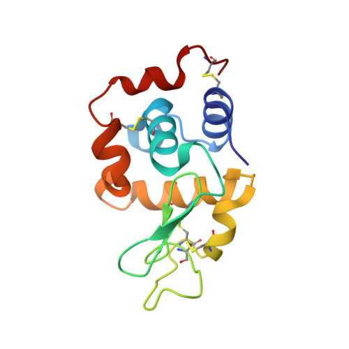
5N12, 6FSJ, 6FSM - PubMed Abstract:
The identification of initial lead conditions for successful protein crystallization is crucial for structural studies using X-ray crystallography. In order to reduce the number of false-negative conditions, an emerging number of fluorescence-based methods have been developed which allow more efficient identification of protein crystals and help to distinguish them from salt crystals. Detection of the native tryptophan fluorescence of protein crystals is one of the most widely used methods. However, this method can fail owing to the properties of the crystallized protein or the chemical composition of the crystallization trials. Here, a simple, fast and cost-efficient method employing 2,2,2-trichloroethanol (TCE) has been developed. It can be performed with a standard UV-light microscope and can be applied to cases in which detection of native tryptophan fluorescence fails. In four test cases this method had no effect on the diffraction properties of the crystals and no structural changes were observed. Further evidence is provided that TCE can be added to crystallization trials during their preparation, making this method compatible with high-throughput approaches.
Organizational Affiliation:
Institute of Biochemistry, University of Cologne, Zülpicher Strasse 47, 50674 Cologne, Germany.