6FLG

Crystal structure of zebrafish Sirtuin 5 in complex with 3(S)-(naphthylthio)succinyl-CPS1 peptide


Experimental Data Snapshot

  • Method: X-RAY DIFFRACTION
  • Resolution: 2.50 Å
  • R-Value Free: 0.311 
  • R-Value Work: 0.242 
  • R-Value Observed: 0.245 

wwPDB Validation   3D Report Full Report


This is version 1.1 of the entry. See complete history


Literature

Potent and Selective Inhibitors of Human Sirtuin 5.

Kalbas, D.Liebscher, S.Nowak, T.Meleshin, M.Pannek, M.Popp, C.Alhalabi, Z.Bordusa, F.Sippl, W.Steegborn, C.Schutkowski, M.

(2018) J Med Chem 61: 2460-2471

  • DOI: https://doi.org/10.1021/acs.jmedchem.7b01648
  • Primary Citation of Related Structures:  
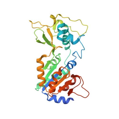
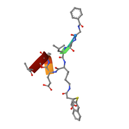
    6FKY, 6FKZ, 6FLG

  • PubMed Abstract: 

    Sirtuins are protein deacylases that regulate metabolism and stress responses and are implicated in aging-related diseases. Modulators of the human sirtuins Sirt1-7 are sought as chemical tools and potential therapeutics, e.g., for cancer. Selective and potent inhibitors are available for Sirt2, but selective inhibitors for Sirt5 with K i values in the low nanomolar range are lacking. We synthesized and screened 3-arylthiosuccinylated and 3-benzylthiosuccinylated peptide derivatives yielding Sirt5 inhibitors with low-nanomolar K i values. A biotinylated derivative with this scaffold represents an affinity probe for human Sirt5 that is able to selectively extract this enzyme out of complex biological samples like cell lysates. Crystal structures of Sirt5/inhibitor complexes reveal that the compounds bind in an unexpected manner to the active site of Sirt5.


  • Organizational Affiliation

    Department of Enzymology, Institute of Biochemistry and Biotechnology , Martin-Luther-University Halle-Wittenberg , 06120 Halle/Saale , Germany.


Macromolecules
Find similar proteins by:  (by identity cutoff)  |  3D Structure
Entity ID: 1
MoleculeChains Sequence LengthOrganismDetailsImage
NAD-dependent protein deacylase sirtuin-5, mitochondrial
A, B
284Danio rerioMutation(s): 0 
Gene Names: sirt5si:ch211-121a2.1
EC: 3.5.1
UniProt
Find proteins for Q6DHI5 (Danio rerio)
Explore Q6DHI5 
Go to UniProtKB:  Q6DHI5
Entity Groups  
Sequence Clusters30% Identity50% Identity70% Identity90% Identity95% Identity100% Identity
UniProt GroupQ6DHI5
Sequence Annotations
Expand
  • Reference Sequence

Find similar proteins by:  Sequence   |   3D Structure  

Entity ID: 2
MoleculeChains Sequence LengthOrganismDetailsImage
3(S)-(naphthylthio)succinyl-CPS1 peptide8Homo sapiensMutation(s): 0 
Sequence Annotations
Expand
  • Reference Sequence
Small Molecules
Ligands 4 Unique
IDChains Name / Formula / InChI Key2D Diagram3D Interactions
EPE
Query on EPE

Download Ideal Coordinates CCD File 
G [auth B]4-(2-HYDROXYETHYL)-1-PIPERAZINE ETHANESULFONIC ACID
C8 H18 N2 O4 S
JKMHFZQWWAIEOD-UHFFFAOYSA-N
DMS
Query on DMS

Download Ideal Coordinates CCD File 
F [auth B]DIMETHYL SULFOXIDE
C2 H6 O S
IAZDPXIOMUYVGZ-UHFFFAOYSA-N
ZN
Query on ZN

Download Ideal Coordinates CCD File 
D [auth A],
H [auth B]
ZINC ION
Zn
PTFCDOFLOPIGGS-UHFFFAOYSA-N
EDO
Query on EDO

Download Ideal Coordinates CCD File 
E [auth A],
I [auth B],
J [auth B]
1,2-ETHANEDIOL
C2 H6 O2
LYCAIKOWRPUZTN-UHFFFAOYSA-N
Experimental Data & Validation

Experimental Data

  • Method: X-RAY DIFFRACTION
  • Resolution: 2.50 Å
  • R-Value Free: 0.311 
  • R-Value Work: 0.242 
  • R-Value Observed: 0.245 
  • Space Group: P 65 2 2
Unit Cell:
Length ( Å )Angle ( ˚ )
a = 87α = 90
b = 87β = 90
c = 316.88γ = 120
Software Package:
Software NamePurpose
REFMACrefinement
XDSdata reduction
XDSdata scaling
PHASERphasing

Structure Validation

View Full Validation Report


Entry History & Funding Information

Deposition Data


Funding OrganizationLocationGrant Number
OberfrankenstiftungGermany04115

Revision History  (Full details and data files)

  • Version 1.0: 2018-04-18
    Type: Initial release
  • Version 1.1: 2024-01-17
    Changes: Data collection, Database references, Derived calculations, Refinement description