6FDQ

Structure of Chlamydia trachomatis effector protein Cdu1 bound to Compound 5


Experimental Data Snapshot

  • Method: X-RAY DIFFRACTION
  • Resolution: 2.30 Å
  • R-Value Free: 0.284 
  • R-Value Work: 0.229 
  • R-Value Observed: 0.232 

wwPDB Validation   3D Report Full Report


Ligand Structure Quality Assessment 


This is version 1.2 of the entry. See complete history


Literature

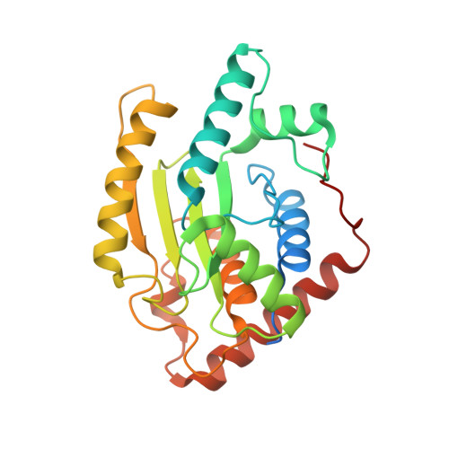
Structural Basis of Substrate Recognition and Covalent Inhibition of Cdu1 from Chlamydia trachomatis.

Ramirez, Y.A.Adler, T.B.Altmann, E.Klemm, T.Tiesmeyer, C.Sauer, F.Kathman, S.G.Statsyuk, A.V.Sotriffer, C.Kisker, C.

(2018) ChemMedChem 13: 2014-2023

  • DOI: https://doi.org/10.1002/cmdc.201800364
  • Primary Citation of Related Structures:  
    6FDK, 6FDQ, 6FDU

  • PubMed Abstract: 

    Based on the similarity between the active sites of the deubiquitylating and deneddylating enzyme ChlaDub1 (Cdu1) and the evolutionarily related protease adenain, a target-hopping screening approach on a focused set of adenain inhibitors was investigated. The cyanopyrimidine-based inhibitors identified represent the first active-site-directed small-molecule inhibitors of Cdu1. High-resolution crystal structures of Cdu1 in complex with two covalently bound cyanopyrimidines, as well as with its substrate ubiquitin, were obtained. These structural data were complemented by enzymatic assays and covalent docking studies to provide insight into the substrate recognition of Cdu1, active-site pocket flexibility and potential hotspots for ligand interaction. Combined, these data provide a strong basis for future structure-guided medicinal chemistry optimization of this cyanopyrimidine scaffold into more potent and selective Cdu1 inhibitors.


  • Organizational Affiliation

    Rudolf Virchow Center for Experimental Biomedicine, Institute of Structural Biology, University of Würzburg, Josef-Schneider-Straße 2, 97080, Würzburg, Germany.


Macromolecules
Find similar proteins by:  (by identity cutoff)  |  3D Structure
Entity ID: 1
MoleculeChains Sequence LengthOrganismDetailsImage
Deubiquitinase and deneddylase Dub1
A, B
266Chlamydia trachomatis L2/434/BuMutation(s): 0 
Gene Names: cdu1CTL0247
EC: 3.4.22
UniProt
Find proteins for B0B9A0 (Chlamydia trachomatis serovar L2 (strain 434/Bu / ATCC VR-902B))
Explore B0B9A0 
Go to UniProtKB:  B0B9A0
Entity Groups  
Sequence Clusters30% Identity50% Identity70% Identity90% Identity95% Identity100% Identity
UniProt GroupB0B9A0
Sequence Annotations
Expand
  • Reference Sequence
Small Molecules
Ligands 1 Unique
IDChains Name / Formula / InChI Key2D Diagram3D Interactions
2UQ
Query on 2UQ

Download Ideal Coordinates CCD File 
C [auth A],
D [auth B]
N-benzyl-2-[(Z)-iminomethyl]pyrimidine-5-carboxamide
C13 H12 N4 O
LOHVEJATQLMRBB-MKMNVTDBSA-N
Experimental Data & Validation

Experimental Data

  • Method: X-RAY DIFFRACTION
  • Resolution: 2.30 Å
  • R-Value Free: 0.284 
  • R-Value Work: 0.229 
  • R-Value Observed: 0.232 
  • Space Group: P 1 21 1
Unit Cell:
Length ( Å )Angle ( ˚ )
a = 43.59α = 90
b = 57.037β = 94.73
c = 114.593γ = 90
Software Package:
Software NamePurpose
REFMACrefinement
XDSdata reduction
Aimlessdata scaling
PHASERphasing

Structure Validation

View Full Validation Report


Ligand Structure Quality Assessment 


Entry History & Funding Information

Deposition Data


Funding OrganizationLocationGrant Number
GSLS University of WuerzburgGermany--

Revision History  (Full details and data files)

  • Version 1.0: 2018-08-15
    Type: Initial release
  • Version 1.1: 2018-10-17
    Changes: Data collection, Database references
  • Version 1.2: 2024-01-17
    Changes: Data collection, Database references, Refinement description