6DL8

Crystal Structure of an influenza A hemagglutinin antibody Fab variant CH67:1203d4 chimera


Experimental Data Snapshot

  • Method: X-RAY DIFFRACTION
  • Resolution: 3.81 Å
  • R-Value Free: 0.284 
  • R-Value Work: 0.233 
  • R-Value Observed: 0.235 

wwPDB Validation   3D Report Full Report


This is version 1.2 of the entry. See complete history


Literature

Identification of Structurally Related Antibodies in Antibody Sequence Databases Using Rosetta-Derived Position-Specific Scoring.

Finn, J.A.Dong, J.Sevy, A.M.Parrish, E.Gilchuk, I.Nargi, R.Scarlett-Jones, M.Reichard, W.Bombardi, R.Voss, T.G.Meiler, J.Crowe Jr., J.E.

(2020) Structure 28: 1124-1130.e5

  • DOI: https://doi.org/10.1016/j.str.2020.07.012
  • Primary Citation of Related Structures:  
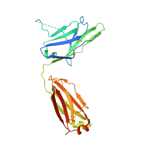
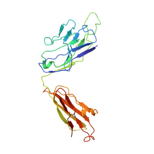
    6DL8, 6DLA, 6DLB

  • PubMed Abstract: 

    The amount of antibody (Ab) variable gene sequence information is expanding rapidly, but our ability to predict the function of Abs from sequence alone is limited. Here, we describe a sequence-to-function prediction method that couples structural data for a single Ab/antigen (Ag) complex with repertoire data. We used a position-specific structure-scoring matrix (P3SM) incorporating structure-prediction scores from Rosetta to identify Ab variable loops that have predicted structural similarity to the influenza virus-specific human Ab CH65. The P3SM approach identified new members of this Ab class. Recombinant Ab expression, crystallography, and virus inhibition assays showed that the HCDR3 loops of the newly identified Abs possessed similar structure and antiviral activity as the comparator CH65. This approach enables discovery of new human Abs with desired structure and function using cDNA repertoires that are obtained readily with current amplicon sequencing techniques.


  • Organizational Affiliation

    Department of Pathology, Microbiology and Immunology, Vanderbilt University Medical Center, Nashville, TN 37232, USA.


Macromolecules
Find similar proteins by:  (by identity cutoff)  |  3D Structure
Entity ID: 1
MoleculeChains Sequence LengthOrganismDetailsImage
CH67:1203d4 FAB light chainA [auth L],
C [auth B]
213Homo sapiensMutation(s): 0 
Entity Groups  
Sequence Clusters30% Identity50% Identity70% Identity90% Identity95% Identity100% Identity
Sequence Annotations
Expand
  • Reference Sequence
Find similar proteins by:  (by identity cutoff)  |  3D Structure
Entity ID: 2
MoleculeChains Sequence LengthOrganismDetailsImage
Ch67:1203d4 FAB heavy chainB [auth H],
D [auth A]
229Homo sapiensMutation(s): 0 
Entity Groups  
Sequence Clusters30% Identity50% Identity70% Identity90% Identity95% Identity100% Identity
Sequence Annotations
Expand
  • Reference Sequence
Experimental Data & Validation

Experimental Data

  • Method: X-RAY DIFFRACTION
  • Resolution: 3.81 Å
  • R-Value Free: 0.284 
  • R-Value Work: 0.233 
  • R-Value Observed: 0.235 
  • Space Group: P 1
Unit Cell:
Length ( Å )Angle ( ˚ )
a = 72.235α = 62.91
b = 73.695β = 88.86
c = 76.848γ = 62.07
Software Package:
Software NamePurpose
PHENIXrefinement
XDSdata reduction
SCALAdata scaling
PHASERphasing

Structure Validation

View Full Validation Report


Entry History 

Deposition Data

Revision History  (Full details and data files)

  • Version 1.0: 2019-06-05
    Type: Initial release
  • Version 1.1: 2020-12-16
    Changes: Database references
  • Version 1.2: 2023-10-11
    Changes: Data collection, Database references, Refinement description