6DHY

Crystallogrpahic tetramer of Zn-bound RIDC1 variant bearing two disulfide bonded cysteines


Experimental Data Snapshot

  • Method: X-RAY DIFFRACTION
  • Resolution: 2.22 Å
  • R-Value Free: 0.258 
  • R-Value Work: 0.229 
  • R-Value Observed: 0.230 

wwPDB Validation   3D Report Full Report


Ligand Structure Quality Assessment 


This is version 2.1 of the entry. See complete history


Literature

Determining the Structural and Energetic Basis of Allostery in a De Novo Designed Metalloprotein Assembly.

Churchfield, L.A.Alberstein, R.G.Williamson, L.M.Tezcan, F.A.

(2018) J Am Chem Soc 140: 10043-10053

  • DOI: https://doi.org/10.1021/jacs.8b05812
  • Primary Citation of Related Structures:  
    6DHY, 6DHZ

  • PubMed Abstract: 

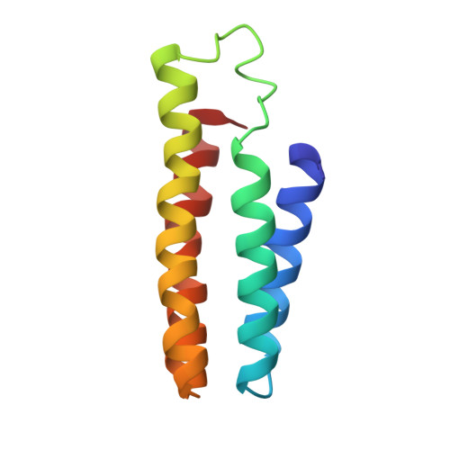
    Despite significant progress in protein design, the construction of protein assemblies that display complex functions (e.g., catalysis or allostery) remains a significant challenge. We recently reported the de novo construction of an allosteric supramolecular protein assembly (Zn- C38/C81/C96 R1 4 ) in which the dissociation and binding of Zn II ions were coupled over a distance of 15 Å to the selective hydrolytic breakage and formation of a single disulfide bond. Zn- C38/C81/C96 R1 4 was constructed by Zn II -templated assembly of a monomeric protein (R1, a derivative of cytochrome cb 562 ) into a tetramer, followed by progressive incorporation of noncovalent and disulfide bonding interactions into the protein-protein interfaces to create a strained quaternary architecture. The interfacial strain thus built allowed mechanical coupling between the binding/dissociation of Zn II and formation/hydrolysis of a single disulfide bond (C38-C38) out of a possible six. While the earlier study provided structural evidence for the two end-states of allosteric coupling, the energetic basis for allosteric coupling and the minimal structural requirements for building this allosteric system were not understood. Toward this end, we have characterized the structures and Zn-binding properties of two related protein constructs ( C38/C96 R1 and C38 R1) which also possess C38-C38 disulfide bonds. In addition, we have carried out extensive molecular dynamics simulations of C38/C81/C96 R1 4 to understand the energetic basis for the selective cleavage of the C38-C38 disulfide bond upon Zn II dissociation. Our analyses reveal that the local interfacial environment around the C38-C38 bond is key to its selective cleavage, but this cleavage is only possible within the context of a stable quaternary architecture which enables structural coupling between Zn II coordination and the protein-protein interfaces.


  • Organizational Affiliation

    Department of Chemistry and Biochemistry , University of California, San Diego , La Jolla , California 92093-0356 , United States.


Macromolecules
Find similar proteins by:  (by identity cutoff)  |  3D Structure
Entity ID: 1
MoleculeChains Sequence LengthOrganismDetailsImage
Soluble cytochrome b562
A, B, C, D
106Escherichia coliMutation(s): 12 
Gene Names: cybC
UniProt
Find proteins for P0ABE7 (Escherichia coli)
Explore P0ABE7 
Go to UniProtKB:  P0ABE7
Entity Groups  
Sequence Clusters30% Identity50% Identity70% Identity90% Identity95% Identity100% Identity
UniProt GroupP0ABE7
Sequence Annotations
Expand
  • Reference Sequence
Small Molecules
Ligands 3 Unique
IDChains Name / Formula / InChI Key2D Diagram3D Interactions
HEC
Query on HEC

Download Ideal Coordinates CCD File 
E [auth A],
I [auth B],
K [auth C],
S [auth D]
HEME C
C34 H34 Fe N4 O4
HXQIYSLZKNYNMH-LJNAALQVSA-N
ZN
Query on ZN

Download Ideal Coordinates CCD File 
G [auth A]
H [auth A]
J [auth B]
N [auth C]
O [auth C]
G [auth A],
H [auth A],
J [auth B],
N [auth C],
O [auth C],
P [auth C],
Q [auth C],
R [auth C],
T [auth D],
U [auth D]
ZINC ION
Zn
PTFCDOFLOPIGGS-UHFFFAOYSA-N
CA
Query on CA

Download Ideal Coordinates CCD File 
F [auth A],
L [auth C],
M [auth C]
CALCIUM ION
Ca
BHPQYMZQTOCNFJ-UHFFFAOYSA-N
Experimental Data & Validation

Experimental Data

  • Method: X-RAY DIFFRACTION
  • Resolution: 2.22 Å
  • R-Value Free: 0.258 
  • R-Value Work: 0.229 
  • R-Value Observed: 0.230 
  • Space Group: P 61
Unit Cell:
Length ( Å )Angle ( ˚ )
a = 52.51α = 90
b = 52.51β = 90
c = 255.67γ = 120
Software Package:
Software NamePurpose
REFMACrefinement
iMOSFLMdata reduction
SCALAdata scaling
PHASERphasing

Structure Validation

View Full Validation Report


Ligand Structure Quality Assessment 


Entry History & Funding Information

Deposition Data


Funding OrganizationLocationGrant Number
National Science Foundation (NSF, United States)United States1607145

Revision History  (Full details and data files)

  • Version 1.0: 2018-07-25
    Type: Initial release
  • Version 1.1: 2018-08-22
    Changes: Data collection, Database references
  • Version 1.2: 2019-11-27
    Changes: Author supporting evidence
  • Version 2.0: 2021-03-10
    Changes: Advisory, Atomic model, Data collection, Derived calculations, Non-polymer description, Structure summary
  • Version 2.1: 2023-10-11
    Changes: Data collection, Database references, Refinement description