6C5T

PPARg LBD bound to SR11023


Experimental Data Snapshot

  • Method: X-RAY DIFFRACTION
  • Resolution: 2.75 Å
  • R-Value Free: 0.286 
  • R-Value Work: 0.254 
  • R-Value Observed: 0.256 

wwPDB Validation   3D Report Full Report


Ligand Structure Quality Assessment 


This is version 1.3 of the entry. See complete history


Literature

PPAR gamma in Complex with an Antagonist and Inverse Agonist: a Tumble and Trap Mechanism of the Activation Helix.

Frkic, R.L.Marshall, A.C.Blayo, A.L.Pukala, T.L.Kamenecka, T.M.Griffin, P.R.Bruning, J.B.

(2018) iScience 5: 69-79

  • DOI: https://doi.org/10.1016/j.isci.2018.06.012
  • Primary Citation of Related Structures:  
    6C5Q, 6C5T

  • PubMed Abstract: 

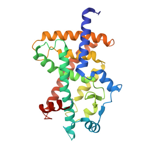
    Peroxisome proliferator activated receptor γ (PPARγ) is a nuclear receptor and target for antidiabetics that increase insulin sensitivity. Owing to the side effects of PPARγ full agonists, research has recently focused on non-activating ligands of PPARγ, which increase insulin sensitivity with decreased side effects. Here, we present the crystal structures of inverse agonist SR10171 and a chemically related antagonist SR11023 bound to the PPARγ ligand-binding domain, revealing an allosteric switch in the activation helix, helix 12 (H12), forming an antagonist conformation in the receptor. H12 interacts with the antagonists to become fixed in an alternative location. Native mass spectrometry indicates that this prevents contacts with coactivator peptides and allows binding of corepressor peptides. Antagonists of related nuclear receptors act to sterically prevent the active configuration of H12, whereas these antagonists of PPARγ alternatively trap H12 in an inactive configuration, which we have termed the tumble and trap mechanism.


  • Organizational Affiliation

    Institute for Photonics and Advanced Sensing (IPAS), School of Biological Sciences, The University of Adelaide, Adelaide, SA 5005, Australia.


Macromolecules
Find similar proteins by:  (by identity cutoff)  |  3D Structure
Entity ID: 1
MoleculeChains Sequence LengthOrganismDetailsImage
Peroxisome proliferator-activated receptor gamma276Homo sapiensMutation(s): 0 
Gene Names: PPARGNR1C3
UniProt & NIH Common Fund Data Resources
Find proteins for P37231 (Homo sapiens)
Explore P37231 
Go to UniProtKB:  P37231
PHAROS:  P37231
GTEx:  ENSG00000132170 
Entity Groups  
Sequence Clusters30% Identity50% Identity70% Identity90% Identity95% Identity100% Identity
UniProt GroupP37231
Sequence Annotations
Expand
  • Reference Sequence
Small Molecules
Ligands 1 Unique
IDChains Name / Formula / InChI Key2D Diagram3D Interactions
EKP
Query on EKP

Download Ideal Coordinates CCD File 
B [auth A]2-{4-[(5-{[(1R)-1-(3-cyclopropylphenyl)ethyl]carbamoyl}-2,3-dimethyl-1H-indol-1-yl)methyl]phenyl}-2-methylpropanoic acid
C33 H36 N2 O3
DVWSHWWLAFDNHD-OAQYLSRUSA-N
Modified Residues  1 Unique
IDChains TypeFormula2D DiagramParent
CSO
Query on CSO
A
L-PEPTIDE LINKINGC3 H7 N O3 SCYS
Experimental Data & Validation

Experimental Data

  • Method: X-RAY DIFFRACTION
  • Resolution: 2.75 Å
  • R-Value Free: 0.286 
  • R-Value Work: 0.254 
  • R-Value Observed: 0.256 
  • Space Group: P 65 2 2
Unit Cell:
Length ( Å )Angle ( ˚ )
a = 63.64α = 90
b = 63.64β = 90
c = 365.35γ = 120
Software Package:
Software NamePurpose
MOSFLMdata reduction
Aimlessdata scaling
PHASERphasing
PHENIXrefinement
PDB_EXTRACTdata extraction

Structure Validation

View Full Validation Report


Ligand Structure Quality Assessment 


Entry History 

Deposition Data

Revision History  (Full details and data files)

  • Version 1.0: 2018-08-01
    Type: Initial release
  • Version 1.1: 2018-09-05
    Changes: Data collection, Database references
  • Version 1.2: 2023-10-04
    Changes: Data collection, Database references, Refinement description
  • Version 1.3: 2023-11-15
    Changes: Data collection