6BB5

Human Oxy-Hemoglobin


Experimental Data Snapshot

  • Method: X-RAY DIFFRACTION
  • Resolution: 2.28 Å
  • R-Value Free: 0.242 
  • R-Value Work: 0.205 
  • R-Value Observed: 0.209 

wwPDB Validation   3D Report Full Report


Ligand Structure Quality Assessment 


This is version 1.1 of the entry. See complete history


Literature

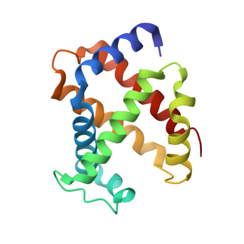
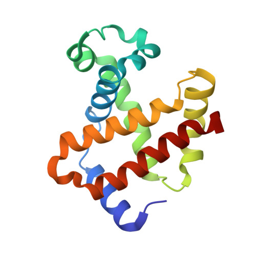
Hemoglobin crystals immersed in liquid oxygen reveal diffusion channels.

Terrell, J.R.Gumpper, R.H.Luo, M.

(2018) Biochem Biophys Res Commun 495: 1858-1863

  • DOI: https://doi.org/10.1016/j.bbrc.2017.12.038
  • Primary Citation of Related Structures:  
    5WOG, 5WOH, 6BB5

  • PubMed Abstract: 

    Human hemoglobin (HbA) transports molecular oxygen (O 2 ) from the lung to tissues where the partial pressure of O 2 is lower. O 2 binds to HbA at the heme cofactor and is stabilized by a distal histidine (HisE7). HisE7 has been observed to occupy opened and closed conformations, and is postulated to act as a gate controlling the binding/release of O 2 . However, it has been suggested that HbA also contains intraprotein oxygen channels for entrances/exits far from the heme. In this study, we developed a novel method of crystal immersion in liquid oxygen prior to X-ray data collection. In the crystals immersed in liquid oxygen, the heme center was oxidized to generate aquomethemoglobin. Increases of structural flexibility were also observed in regions that are synonymous with previously postulated oxygen channels. These regions also correspond to medically relevant mutations which affect O 2 affinity. The way HbA utilizes these O 2 channels could have a profound impact on understanding the relationship of HbA O 2 transport within these disease conditions. Finally, the liquid oxygen immersion technique can be utilized as a new tool to crystallographically examine proteins and protein complexes which utilize O 2 for enzyme catalysis or transport.


  • Organizational Affiliation

    Department of Chemistry, Georgia State University, Atlanta, GA 30302, USA; Center for Diagnostics and Therapeutics, Georgia State University, Atlanta, GA 30302, USA.


Macromolecules
Find similar proteins by:  (by identity cutoff)  |  3D Structure
Entity ID: 1
MoleculeChains Sequence LengthOrganismDetailsImage
Hemoglobin subunit alpha139Homo sapiensMutation(s): 0 
UniProt & NIH Common Fund Data Resources
Find proteins for P69905 (Homo sapiens)
Explore P69905 
Go to UniProtKB:  P69905
PHAROS:  P69905
Entity Groups  
Sequence Clusters30% Identity50% Identity70% Identity90% Identity95% Identity100% Identity
UniProt GroupP69905
Sequence Annotations
Expand
  • Reference Sequence
Find similar proteins by:  (by identity cutoff)  |  3D Structure
Entity ID: 2
MoleculeChains Sequence LengthOrganismDetailsImage
Hemoglobin subunit beta145Homo sapiensMutation(s): 0 
UniProt & NIH Common Fund Data Resources
Find proteins for P68871 (Homo sapiens)
Explore P68871 
Go to UniProtKB:  P68871
PHAROS:  P68871
GTEx:  ENSG00000244734 
Entity Groups  
Sequence Clusters30% Identity50% Identity70% Identity90% Identity95% Identity100% Identity
UniProt GroupP68871
Sequence Annotations
Expand
  • Reference Sequence
Experimental Data & Validation

Experimental Data

  • Method: X-RAY DIFFRACTION
  • Resolution: 2.28 Å
  • R-Value Free: 0.242 
  • R-Value Work: 0.205 
  • R-Value Observed: 0.209 
  • Space Group: P 41 21 2
Unit Cell:
Length ( Å )Angle ( ˚ )
a = 53.73α = 90
b = 53.73β = 90
c = 192.29γ = 90
Software Package:
Software NamePurpose
XSCALEdata scaling
PHENIXrefinement
PDB_EXTRACTdata extraction
XDSdata reduction
PHENIXphasing

Structure Validation

View Full Validation Report


Ligand Structure Quality Assessment 


Entry History 

Deposition Data

Revision History  (Full details and data files)

  • Version 1.0: 2018-01-10
    Type: Initial release
  • Version 1.1: 2023-10-04
    Changes: Data collection, Database references, Derived calculations, Refinement description