


X-ray Crystal Structure of Pseudoazurin Met16Leu Variant
Sakai, C., Yamaguchi, T., Kohzuma, T.To be published.
Experimental Data Snapshot
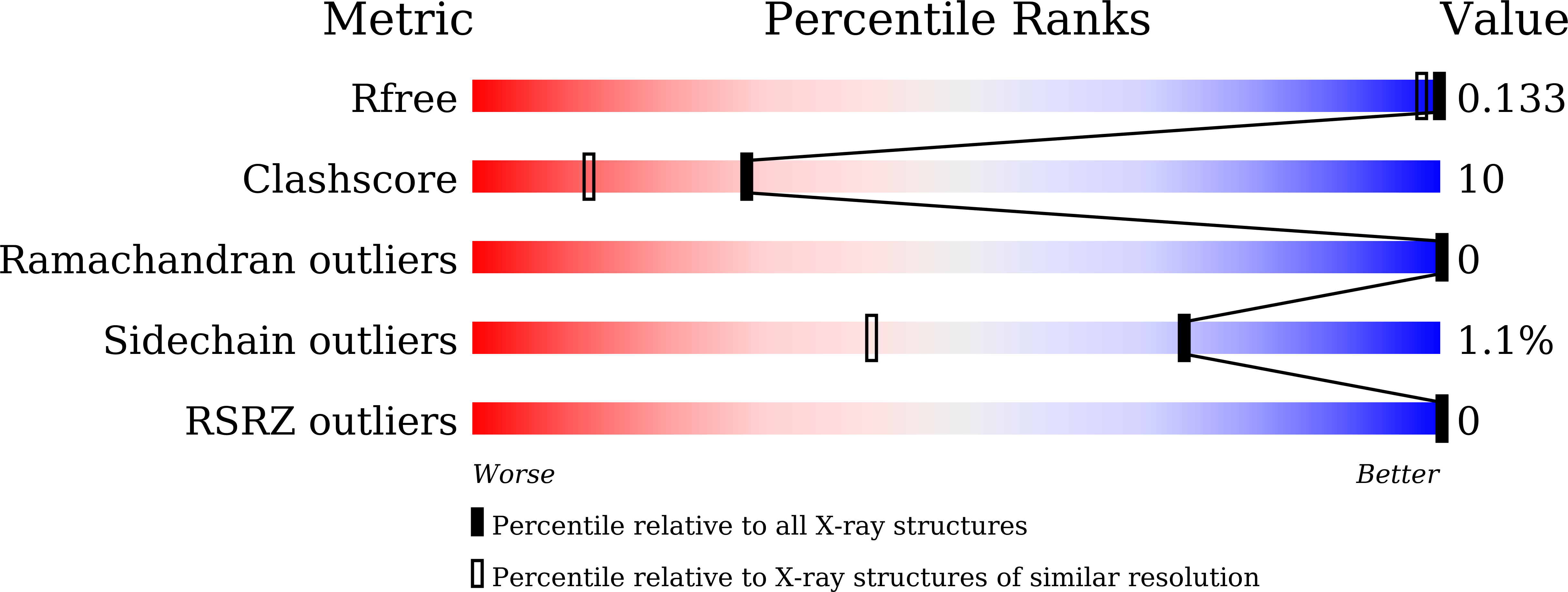
wwPDB Validation 3D Report Full Report
Entity ID: 1 | |||||
|---|---|---|---|---|---|
| Molecule | Chains | Sequence Length | Organism | Details | Image |
| Pseudoazurin | 124 | Achromobacter cycloclastes | Mutation(s): 1 Gene Names: bcp | 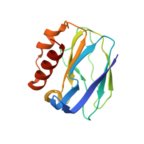 | |
UniProt | |||||
Find proteins for P19567 (Achromobacter cycloclastes) Explore P19567 Go to UniProtKB: P19567 | |||||
Entity Groups | |||||
| Sequence Clusters | 30% Identity50% Identity70% Identity90% Identity95% Identity100% Identity | ||||
| UniProt Group | P19567 | ||||
Sequence AnnotationsExpand | |||||
| |||||
| Ligands 1 Unique | |||||
|---|---|---|---|---|---|
| ID | Chains | Name / Formula / InChI Key | 2D Diagram | 3D Interactions | |
| CU Query on CU | C [auth A], D [auth B] | COPPER (II) ION Cu JPVYNHNXODAKFH-UHFFFAOYSA-N |  | ||
| Length ( Å ) | Angle ( ˚ ) |
|---|---|
| a = 34.05 | α = 90 |
| b = 60.22 | β = 95.8 |
| c = 48.03 | γ = 90 |
| Software Name | Purpose |
|---|---|
| SHELX | refinement |
| SCALA | data scaling |
| PDB_EXTRACT | data extraction |
| iMOSFLM | data reduction |
| MOLREP | phasing |
| Funding Organization | Location | Grant Number |
|---|---|---|
| Japan Society for the Promotion of Science | Japan | 22550145 |
RCSB PDB (citation) is hosted by
RCSB PDB is a member of the

