6A97

Crystal structure of MHC-like MILL2


Experimental Data Snapshot

  • Method: X-RAY DIFFRACTION
  • Resolution: 2.15 Å
  • R-Value Free: 0.245 
  • R-Value Work: 0.213 
  • R-Value Observed: 0.214 

wwPDB Validation   3D Report Full Report


This is version 1.0 of the entry. See complete history


Literature

Structure of MHC class I-like MILL2 reveals heparan-sulfate binding and interdomain flexibility.

Kajikawa, M.Ose, T.Fukunaga, Y.Okabe, Y.Matsumoto, N.Yonezawa, K.Shimizu, N.Kollnberger, S.Kasahara, M.Maenaka, K.

(2018) Nat Commun 9: 4330-4330

  • DOI: https://doi.org/10.1038/s41467-018-06797-8
  • Primary Citation of Related Structures:  
    6A97

  • PubMed Abstract: 

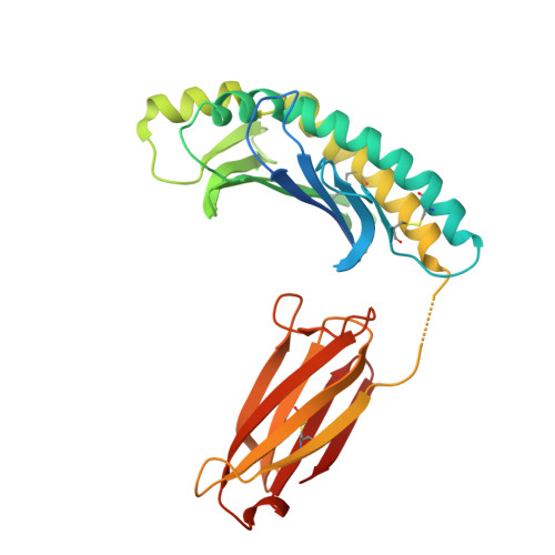
    The MILL family, composed of MILL1 and MILL2, is a group of nonclassical MHC class I molecules that occur in some orders of mammals. It has been reported that mouse MILL2 is involved in wound healing; however, the molecular mechanisms remain unknown. Here, we determine the crystal structure of MILL2 at 2.15 Å resolution, revealing an organization similar to classical MHC class I. However, the α1-α2 domains are not tightly fixed on the α3-β 2 m domains, indicating unusual interdomain flexibility. The groove between the two helices in the α1-α2 domains is too narrow to permit ligand binding. Notably, an unusual basic patch on the α3 domain is involved in the binding to heparan sulfate which is essential for MILL2 interactions with fibroblasts. These findings suggest that MILL2 has a unique structural architecture and physiological role, with binding to heparan sulfate proteoglycans on fibroblasts possibly regulating cellular recruitment in biological events.


  • Organizational Affiliation

    Laboratory of Microbiology, Showa Pharmaceutical University, Machida, Tokyo, 190-8543, Japan.


Macromolecules
Find similar proteins by:  (by identity cutoff)  |  3D Structure
Entity ID: 1
MoleculeChains Sequence LengthOrganismDetailsImage
MHC I-like leukocyte 2 long form
A, C
292Mus musculusMutation(s): 0 
Gene Names: Mill2
UniProt
Find proteins for Q8HWE5 (Mus musculus)
Explore Q8HWE5 
Go to UniProtKB:  Q8HWE5
Entity Groups  
Sequence Clusters30% Identity50% Identity70% Identity90% Identity95% Identity100% Identity
UniProt GroupQ8HWE5
Sequence Annotations
Expand
  • Reference Sequence
Find similar proteins by:  (by identity cutoff)  |  3D Structure
Entity ID: 2
MoleculeChains Sequence LengthOrganismDetailsImage
Beta-2-microglobulin
B, D
100Mus musculusMutation(s): 0 
Gene Names: B2m
UniProt & NIH Common Fund Data Resources
Find proteins for P01887 (Mus musculus)
Explore P01887 
Go to UniProtKB:  P01887
IMPC:  MGI:88127
Entity Groups  
Sequence Clusters30% Identity50% Identity70% Identity90% Identity95% Identity100% Identity
UniProt GroupP01887
Sequence Annotations
Expand
  • Reference Sequence
Small Molecules
Ligands 1 Unique
IDChains Name / Formula / InChI Key2D Diagram3D Interactions
SO4
Query on SO4

Download Ideal Coordinates CCD File 
E [auth C]SULFATE ION
O4 S
QAOWNCQODCNURD-UHFFFAOYSA-L
Experimental Data & Validation

Experimental Data

  • Method: X-RAY DIFFRACTION
  • Resolution: 2.15 Å
  • R-Value Free: 0.245 
  • R-Value Work: 0.213 
  • R-Value Observed: 0.214 
  • Space Group: P 21 21 21
Unit Cell:
Length ( Å )Angle ( ˚ )
a = 89.633α = 90
b = 93.497β = 90
c = 138.123γ = 90
Software Package:
Software NamePurpose
PHENIXrefinement
HKL-2000data reduction
HKL-2000data scaling
MOLREPphasing

Structure Validation

View Full Validation Report


Entry History & Funding Information

Deposition Data


Funding OrganizationLocationGrant Number
Ministry of Education, Culture, Sports, Science and Technology (Japan)Japan20057020

Revision History  (Full details and data files)

  • Version 1.0: 2018-12-05
    Type: Initial release