5ZXM

Crystal Structure of GyraseB N-terminal at 1.93A Resolution


Experimental Data Snapshot

  • Method: X-RAY DIFFRACTION
  • Resolution: 1.94 Å
  • R-Value Free: 0.226 
  • R-Value Work: 0.181 
  • R-Value Observed: 0.182 

wwPDB Validation   3D Report Full Report


Ligand Structure Quality Assessment 


This is version 2.2 of the entry. See complete history


Literature

Structural insights into the transient closed conformation and pH dependent ATPase activity of S.Typhi GyraseB N- terminal domain.

Gupta, D.Tiwari, P.Haque, M.A.Sachdeva, E.Hassan, M.I.Ethayathulla, A.S.Kaur, P.

(2021) Arch Biochem Biophys 701: 108786-108786

  • DOI: https://doi.org/10.1016/j.abb.2021.108786

  • PubMed Abstract: 

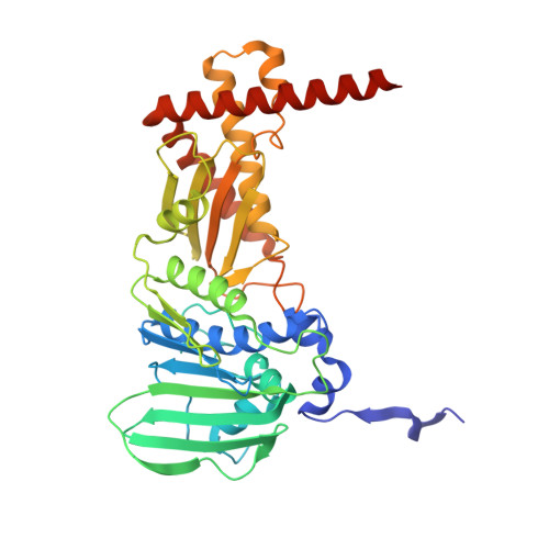
    DNA Gyrase is a type II topoisomerase that utilizes the energy of ATP hydrolysis for introducing negative supercoils in DNA. The protein comprises two subunits GyrA and GyrB that form a GyrA 2 GyrB 2 heterotetramer. GyrB subunit contains the N-terminal domain (GBNTD) for ATPase activity and the C-terminal domain (GBCTD) for interaction with GyrA and DNA. Earlier structural studies have revealed three different conformational states for GBNTD during ATP hydrolysis defined as open, semi-open, and closed. Here we report, the three-dimensional structure of a new transient closed conformation of GBNTD from Salmonella Typhi (StGBNTD) at 1.94 Å resolution. Based on the structural analysis of this transient closed conformation, we propose the role of protein in the mechanism of ATP hydrolysis. We further explored the effect of pH on ATPase activity and structural stability of the GBNTD using CD and fluorescence spectroscopy at varying pH environment. Kinetic parameters obtained from the ATPase assay were correlated with its secondary and tertiary structure at their respective pH environment. The protein possessed maximum ATPase activity and structural stability at optimum pH 8. At acidic pH, a remarkable decrease in both enzymatic activity and structural stability was observed whereas at alkaline pH there was no significant change. The structural analysis of StGBNTD reveals the role of polar interactions in stabilizing the overall dimeric conformation of the protein.


  • Organizational Affiliation

    Department of Biophysics, All India Institute of Medical Sciences, Ansari Nagar, New Delhi, 110029, India.


Macromolecules
Find similar proteins by:  (by identity cutoff)  |  3D Structure
Entity ID: 1
MoleculeChains Sequence LengthOrganismDetailsImage
DNA gyrase subunit B
A, B
400Salmonella enterica subsp. entericaMutation(s): 4 
Gene Names: gyrB
EC: 5.6.2.2
UniProt
Find proteins for A0A3R0U328 (Salmonella enterica I)
Explore A0A3R0U328 
Go to UniProtKB:  A0A3R0U328
Entity Groups  
Sequence Clusters30% Identity50% Identity70% Identity90% Identity95% Identity100% Identity
UniProt GroupA0A3R0U328
Sequence Annotations
Expand
  • Reference Sequence
Small Molecules
Ligands 5 Unique
IDChains Name / Formula / InChI Key2D Diagram3D Interactions
ADP
Query on ADP

Download Ideal Coordinates CCD File 
C [auth A],
H [auth B]
ADENOSINE-5'-DIPHOSPHATE
C10 H15 N5 O10 P2
XTWYTFMLZFPYCI-KQYNXXCUSA-N
PEG
Query on PEG

Download Ideal Coordinates CCD File 
E [auth A],
F [auth A],
J [auth B]
DI(HYDROXYETHYL)ETHER
C4 H10 O3
MTHSVFCYNBDYFN-UHFFFAOYSA-N
SO4
Query on SO4

Download Ideal Coordinates CCD File 
D [auth A],
I [auth B]
SULFATE ION
O4 S
QAOWNCQODCNURD-UHFFFAOYSA-L
GOL
Query on GOL

Download Ideal Coordinates CCD File 
K [auth B],
L [auth B]
GLYCEROL
C3 H8 O3
PEDCQBHIVMGVHV-UHFFFAOYSA-N
MG
Query on MG

Download Ideal Coordinates CCD File 
G [auth A]MAGNESIUM ION
Mg
JLVVSXFLKOJNIY-UHFFFAOYSA-N
Experimental Data & Validation

Experimental Data

  • Method: X-RAY DIFFRACTION
  • Resolution: 1.94 Å
  • R-Value Free: 0.226 
  • R-Value Work: 0.181 
  • R-Value Observed: 0.182 
  • Space Group: P 2 21 21
Unit Cell:
Length ( Å )Angle ( ˚ )
a = 79.42α = 90
b = 86.53β = 90
c = 135.99γ = 90
Software Package:
Software NamePurpose
REFMACrefinement
XDSdata reduction
autoPROCdata scaling
PHASERphasing

Structure Validation

View Full Validation Report


Ligand Structure Quality Assessment 


Entry History 

Deposition Data

Revision History  (Full details and data files)

  • Version 1.0: 2019-05-22
    Type: Initial release
  • Version 2.0: 2020-07-15
    Type: Coordinate replacement
    Reason: Model orientation/position
    Changes: Advisory, Atomic model, Data collection, Database references, Derived calculations, Refinement description, Source and taxonomy, Structure summary
  • Version 2.1: 2021-03-10
    Changes: Database references, Refinement description
  • Version 2.2: 2023-11-22
    Changes: Data collection, Database references, Refinement description