Protein crystal quality oriented disulfide bond engineering.
Pu, M., Xu, Z., Peng, Y., Hou, Y., Liu, D., Wang, Y., Liu, H., Song, G., Liu, Z.J.(2018) Protein Cell 9: 659-663
- PubMed: 29039033
- DOI: https://doi.org/10.1007/s13238-017-0482-7
- Primary Citation of Related Structures:
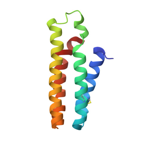
5YM7, 5YO3, 5YO4, 5YO5, 5YO6, 5YOB, 5YOC, 5YOE, 5YOG
Organizational Affiliation:
National Laboratory of Biomacromolecules, Institute of Biophysics, Chinese Academy of Sciences, Beijing, 100101, China.