


X-ray crystal structure of Pseudoazurin Met16Phe variant
Yamaguchi, T., Akao, K., Kohzuma, T.To be published.
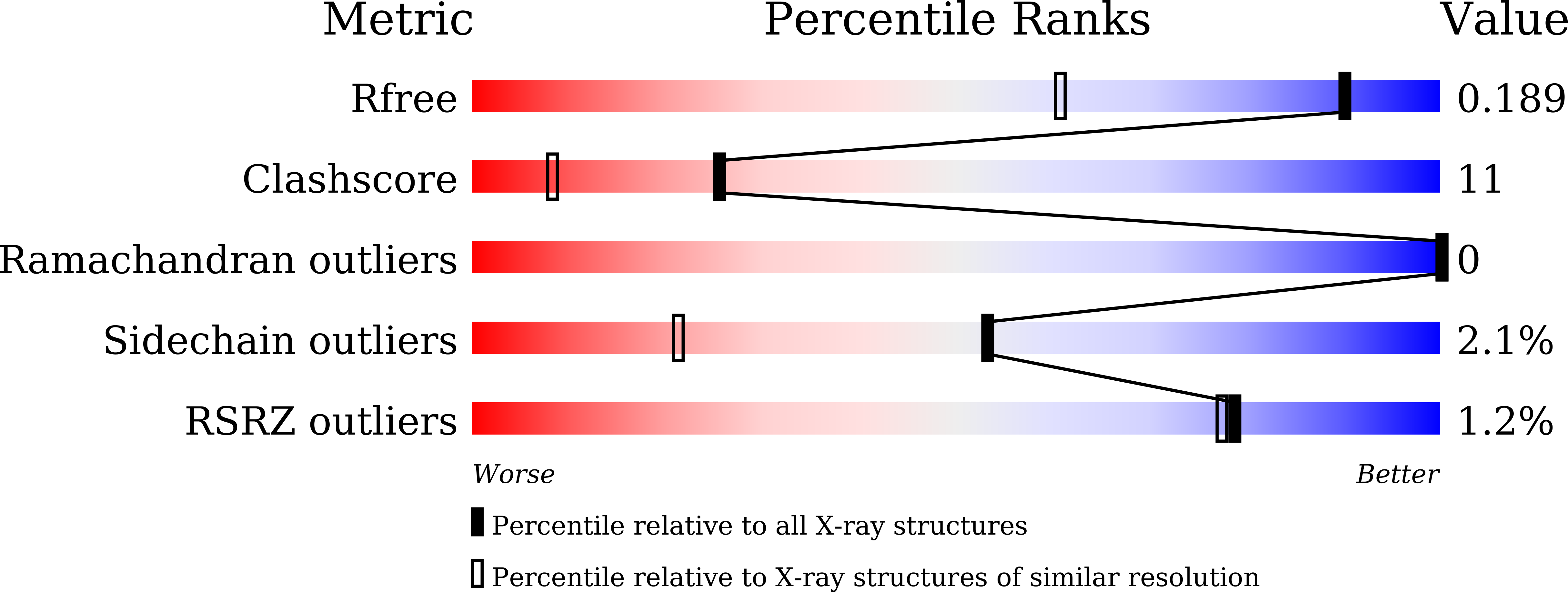
Experimental Data Snapshot
wwPDB Validation 3D Report Full Report
Entity ID: 1 | |||||
|---|---|---|---|---|---|
| Molecule | Chains | Sequence Length | Organism | Details | Image |
| Pseudoazurin | 124 | Achromobacter cycloclastes | Mutation(s): 1 Gene Names: bcp | 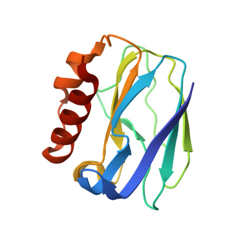 | |
UniProt | |||||
Find proteins for P19567 (Achromobacter cycloclastes) Explore P19567 Go to UniProtKB: P19567 | |||||
Entity Groups | |||||
| Sequence Clusters | 30% Identity50% Identity70% Identity90% Identity95% Identity100% Identity | ||||
| UniProt Group | P19567 | ||||
Sequence AnnotationsExpand | |||||
| |||||
| Ligands 2 Unique | |||||
|---|---|---|---|---|---|
| ID | Chains | Name / Formula / InChI Key | 2D Diagram | 3D Interactions | |
| GOL Query on GOL | D [auth A] E [auth A] F [auth A] H [auth B] I [auth B] | GLYCEROL C3 H8 O3 PEDCQBHIVMGVHV-UHFFFAOYSA-N |  | ||
| CU (Subject of Investigation/LOI) Query on CU | C [auth A], G [auth B] | COPPER (II) ION Cu JPVYNHNXODAKFH-UHFFFAOYSA-N |  | ||
| Length ( Å ) | Angle ( ˚ ) |
|---|---|
| a = 34.715 | α = 90 |
| b = 59.62 | β = 105.37 |
| c = 54.324 | γ = 90 |
| Software Name | Purpose |
|---|---|
| REFMAC | refinement |
| MOLREP | phasing |
RCSB PDB (citation) is hosted by
RCSB PDB is a member of the

