5X41

3.5A resolution structure of a cobalt energy-coupling factor transporter using LCP method-CbiMQO


Experimental Data Snapshot

  • Method: X-RAY DIFFRACTION
  • Resolution: 3.47 Å
  • R-Value Free: 0.335 
  • R-Value Work: 0.299 
  • R-Value Observed: 0.300 

wwPDB Validation   3D Report Full Report


This is version 1.3 of the entry. See complete history


Literature

Structure and mechanism of a group-I cobalt energy coupling factor transporter

Bao, Z.Qi, X.Hong, S.Xu, K.He, F.Zhang, M.Chen, J.Chao, D.Zhao, W.Li, D.Wang, J.Zhang, P.

(2017) Cell Res 27: 675-687

  • DOI: https://doi.org/10.1038/cr.2017.38
  • Primary Citation of Related Structures:  
    5X3X, 5X40, 5X41

  • PubMed Abstract: 

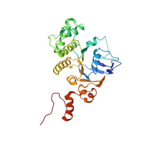
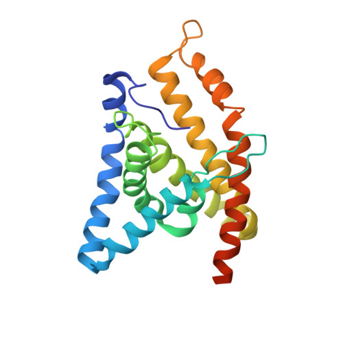
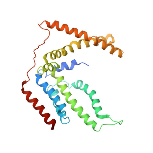
    Energy-coupling factor (ECF) transporters are a large family of ATP-binding cassette transporters recently identified in microorganisms. Responsible for micronutrient uptake from the environment, ECF transporters are modular transporters composed of a membrane substrate-binding component EcfS and an ECF module consisting of an integral membrane scaffold component EcfT and two cytoplasmic ATP binding/hydrolysis components EcfA/A'. ECF transporters are classified into groups I and II. Currently, the molecular understanding of group-I ECF transporters is very limited, partly due to a lack of transporter complex structural information. Here, we present structures and structure-based analyses of the group-I cobalt ECF transporter CbiMNQO, whose constituting subunits CbiM/CbiN, CbiQ, and CbiO correspond to the EcfS, EcfT, and EcfA components of group-II ECF transporters, respectively. Through reconstitution of different CbiMNQO subunits and determination of related ATPase and transporter activities, the substrate-binding subunit CbiM was found to stimulate CbiQO's basal ATPase activity. The structure of CbiMQO complex was determined in its inward-open conformation and that of CbiO in β, γ-methyleneadenosine 5'-triphosphate-bound closed conformation. Structure-based analyses revealed interactions between different components, substrate-gating function of the L1 loop of CbiM, and conformational changes of CbiO induced by ATP binding and product release within the CbiMNQO transporter complex. These findings enabled us to propose a working model of the CbiMNQO transporter, in which the transport process requires the rotation or toppling of both CbiQ and CbiM, and CbiN might function in coupling conformational changes between CbiQ and CbiM.


  • Organizational Affiliation

    National Key Laboratory of Plant Molecular Genetics, CAS Center for Excellence in Molecular Plant Sciences, Institute of Plant Physiology and Ecology, Shanghai Institutes for Biological Sciences, Chinese Academy of Sciences, Shanghai 200032, China.


Macromolecules
Find similar proteins by:  (by identity cutoff)  |  3D Structure
Entity ID: 1
MoleculeChains Sequence LengthOrganismDetailsImage
Cobalt ABC transporter ATP-binding proteinA,
B,
E [auth C],
F [auth D]
280Rhodobacter capsulatusMutation(s): 0 
Membrane Entity: Yes 
UniProt
Find proteins for A0A1G7HZ57 (Rhodobacter capsulatus)
Explore A0A1G7HZ57 
Go to UniProtKB:  A0A1G7HZ57
Entity Groups  
Sequence Clusters30% Identity50% Identity70% Identity90% Identity95% Identity100% Identity
UniProt GroupA0A1G7HZ57
Sequence Annotations
Expand
  • Reference Sequence
Find similar proteins by:  (by identity cutoff)  |  3D Structure
Entity ID: 2
MoleculeChains Sequence LengthOrganismDetailsImage
Cobalt transport protein CbiMC [auth M],
G [auth E]
222Rhodobacter capsulatusMutation(s): 0 
Gene Names: cbiM
Membrane Entity: Yes 
UniProt
Find proteins for A0A0Q0R232 (Rhodobacter capsulatus)
Explore A0A0Q0R232 
Go to UniProtKB:  A0A0Q0R232
Entity Groups  
Sequence Clusters30% Identity50% Identity70% Identity90% Identity95% Identity100% Identity
UniProt GroupA0A0Q0R232
Sequence Annotations
Expand
  • Reference Sequence
Find similar proteins by:  (by identity cutoff)  |  3D Structure
Entity ID: 3
MoleculeChains Sequence LengthOrganismDetailsImage
Uncharacterized protein CbiQD [auth Q],
H [auth F]
256Rhodobacter capsulatusMutation(s): 0 
Membrane Entity: Yes 
UniProt
Find proteins for A0A1G7HYD7 (Rhodobacter capsulatus)
Explore A0A1G7HYD7 
Go to UniProtKB:  A0A1G7HYD7
Entity Groups  
Sequence Clusters30% Identity50% Identity70% Identity90% Identity95% Identity100% Identity
UniProt GroupA0A1G7HYD7
Sequence Annotations
Expand
  • Reference Sequence
Experimental Data & Validation

Experimental Data

  • Method: X-RAY DIFFRACTION
  • Resolution: 3.47 Å
  • R-Value Free: 0.335 
  • R-Value Work: 0.299 
  • R-Value Observed: 0.300 
  • Space Group: P 21 21 21
Unit Cell:
Length ( Å )Angle ( ˚ )
a = 96.159α = 90
b = 135.04β = 90
c = 204.424γ = 90
Software Package:
Software NamePurpose
PHENIXrefinement
HKL-3000data collection
HKL-3000data processing

Structure Validation

View Full Validation Report


Entry History 

Deposition Data

Revision History  (Full details and data files)

  • Version 1.0: 2017-04-19
    Type: Initial release
  • Version 1.1: 2017-05-17
    Changes: Database references
  • Version 1.2: 2017-10-04
    Changes: Data collection
  • Version 1.3: 2024-03-27
    Changes: Data collection, Database references