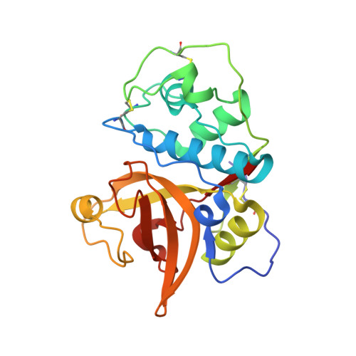
Crystal Structures Of Mite Allergens Der F 1 And Der P 1 Reveal Differences In Surface-Exposed Residues That May Influence Antibody Binding
Chruszcz, M., Chapman, M.D., Vailes, L.D., Stura, E.A., Saint-Remy, J.M., Minor, W., Pomes, A.(2009) J Mol Biol 386: 520
- PubMed: 19136006
- DOI: https://doi.org/10.1016/j.jmb.2008.12.049
- Primary Citation of Related Structures:
3F5V, 5VPK - PubMed Abstract:
The group 1 mite allergens Der f 1 and Der p 1 are potent allergens excreted by Dermatophagoides farinae and Dermatophagoides pteronyssinus, respectively. The human immunoglobulin E antibody responses to the group 1 allergens show more cross-reactivity than the murine immunoglobulin G antibody responses, which are largely species specific. Here, we report the crystal structure of the mature form of Der f 1, which was isolated from its natural source, and a new high-resolution structure of mature recombinant Der p 1. Unlike Der p 1, Der f 1 is monomeric both in the crystalline state and in solution. Moreover, no metal binding is observed in the structure of Der f 1 despite the fact that all amino acids involved in Ca(2+) binding in Der p 1 are completely conserved in Der f 1. Although Der p 1 and Der f 1 share an extensive sequence identity, comparison of the crystal structures of both allergens revealed structural features that could explain the differences in murine IgG and human IgE antibody responses to these allergens. There are structural differences between Der f 1 and Der p 1 that are unevenly distributed on the allergens' surfaces. This uneven spatial arrangement of conserved versus altered residues could explain both the specificity and cross-reactivity of antibodies against Der f 1 and Der p 1.
Organizational Affiliation:
Department of Molecular Physiology and Biological Physics, University of Virginia, Charlottesville, 22908, USA.