5T04

STRUCTURE OF CONSTITUTIVELY ACTIVE NEUROTENSIN RECEPTOR


Experimental Data Snapshot

  • Method: X-RAY DIFFRACTION
  • Resolution: 3.30 Å
  • R-Value Free: 0.283 
  • R-Value Work: 0.253 
  • R-Value Observed: 0.256 

wwPDB Validation   3D Report Full Report


Ligand Structure Quality Assessment 


This is version 1.1 of the entry. See complete history


Literature

Structure and dynamics of a constitutively active neurotensin receptor.

Krumm, B.E.Lee, S.Bhattacharya, S.Botos, I.White, C.F.Du, H.Vaidehi, N.Grisshammer, R.

(2016) Sci Rep 6: 38564-38564

  • DOI: https://doi.org/10.1038/srep38564
  • Primary Citation of Related Structures:  
    5T04

  • PubMed Abstract: 

    Many G protein-coupled receptors show constitutive activity, resulting in the production of a second messenger in the absence of an agonist; and naturally occurring constitutively active mutations in receptors have been implicated in diseases. To gain insight into mechanistic aspects of constitutive activity, we report here the 3.3 Å crystal structure of a constitutively active, agonist-bound neurotensin receptor (NTSR1) and molecular dynamics simulations of agonist-occupied and ligand-free receptor. Comparison with the structure of a NTSR1 variant that has little constitutive activity reveals uncoupling of the ligand-binding domain from conserved connector residues, that effect conformational changes during GPCR activation. Furthermore, molecular dynamics simulations show strong contacts between connector residue side chains and increased flexibility at the intracellular receptor face as features that coincide with robust signalling in cells. The loss of correlation between the binding pocket and conserved connector residues, combined with altered receptor dynamics, possibly explains the reduced neurotensin efficacy in the constitutively active NTSR1 and a facilitated initial engagement with G protein in the absence of agonist.


  • Organizational Affiliation

    Membrane Protein Structure Function Unit, National Institute of Neurological Disorders and Stroke, National Institutes of Health, Department of Health and Human Services, Rockville, Maryland 20852, United States.


Macromolecules
Find similar proteins by:  (by identity cutoff)  |  3D Structure
Entity ID: 1
MoleculeChains Sequence LengthOrganismDetailsImage
Neurotensin receptor type 1,Endolysin,Neurotensin receptor type 1513Rattus norvegicusTequatrovirus T4Mutation(s): 10 
Gene Names: Ntsr1Ntsr
EC: 3.2.1.17
Membrane Entity: Yes 
UniProt
Find proteins for P20789 (Rattus norvegicus)
Explore P20789 
Go to UniProtKB:  P20789
Find proteins for P00720 (Enterobacteria phage T4)
Explore P00720 
Go to UniProtKB:  P00720
Entity Groups  
Sequence Clusters30% Identity50% Identity70% Identity90% Identity95% Identity100% Identity
UniProt GroupsP20789P00720
Sequence Annotations
Expand
  • Reference Sequence

Find similar proteins by:  Sequence   |   3D Structure  

Entity ID: 2
MoleculeChains Sequence LengthOrganismDetailsImage
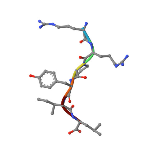
ARG-ARG-PRO-TYR-ILE-LEU6Rattus norvegicusMutation(s): 0 
Membrane Entity: Yes 
UniProt
Find proteins for P20068 (Rattus norvegicus)
Explore P20068 
Go to UniProtKB:  P20068
Entity Groups  
UniProt GroupP20068
Sequence Annotations
Expand
  • Reference Sequence
Experimental Data & Validation

Experimental Data

  • Method: X-RAY DIFFRACTION
  • Resolution: 3.30 Å
  • R-Value Free: 0.283 
  • R-Value Work: 0.253 
  • R-Value Observed: 0.256 
  • Space Group: P 21 21 2
Unit Cell:
Length ( Å )Angle ( ˚ )
a = 104.19α = 90
b = 75.71β = 90
c = 83.2γ = 90
Software Package:
Software NamePurpose
PHENIXrefinement
XDSdata reduction
XDSdata scaling
PHASERphasing

Structure Validation

View Full Validation Report


Ligand Structure Quality Assessment 


Entry History 

Deposition Data

Revision History  (Full details and data files)

  • Version 1.0: 2016-12-21
    Type: Initial release
  • Version 1.1: 2023-10-04
    Changes: Data collection, Database references, Refinement description