5OXI

C-terminally retracted ubiquitin L67S mutant


Experimental Data Snapshot

  • Method: X-RAY DIFFRACTION
  • Resolution: 1.63 Å
  • R-Value Free: 0.237 
  • R-Value Work: 0.193 
  • R-Value Observed: 0.196 

wwPDB Validation   3D Report Full Report


This is version 1.2 of the entry. See complete history


Literature

An invisible ubiquitin conformation is required for efficient phosphorylation by PINK1.

Gladkova, C.Schubert, A.F.Wagstaff, J.L.Pruneda, J.N.Freund, S.M.Komander, D.

(2017) EMBO J 36: 3555-3572

  • DOI: https://doi.org/10.15252/embj.201797876
  • Primary Citation of Related Structures:  
    5OXH, 5OXI

  • PubMed Abstract: 

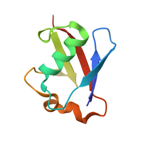
    The Ser/Thr protein kinase PINK1 phosphorylates the well-folded, globular protein ubiquitin (Ub) at a relatively protected site, Ser65. We previously showed that Ser65 phosphorylation results in a conformational change in which Ub adopts a dynamic equilibrium between the known, common Ub conformation and a distinct, second conformation wherein the last β-strand is retracted to extend the Ser65 loop and shorten the C-terminal tail. We show using chemical exchange saturation transfer (CEST) nuclear magnetic resonance experiments that a similar, C-terminally retracted (Ub-CR) conformation also exists at low population in wild-type Ub. Point mutations in the moving β5 and neighbouring β-strands shift the Ub/Ub-CR equilibrium. This enabled functional studies of the two states, and we show that while the Ub-CR conformation is defective for conjugation, it demonstrates improved binding to PINK1 through its extended Ser65 loop, and is a superior PINK1 substrate. Together our data suggest that PINK1 utilises a lowly populated yet more suitable Ub-CR conformation of Ub for efficient phosphorylation. Our findings could be relevant for many kinases that phosphorylate residues in folded protein domains.


  • Organizational Affiliation

    Medical Research Council Laboratory of Molecular Biology, Cambridge, UK.


Macromolecules
Find similar proteins by:  (by identity cutoff)  |  3D Structure
Entity ID: 1
MoleculeChains Sequence LengthOrganismDetailsImage
Ubiquitin L67S mutant
A, B
76Homo sapiensMutation(s): 1 
UniProt & NIH Common Fund Data Resources
Find proteins for P0CG48 (Homo sapiens)
Explore P0CG48 
Go to UniProtKB:  P0CG48
PHAROS:  P0CG48
GTEx:  ENSG00000150991 
Entity Groups  
Sequence Clusters30% Identity50% Identity70% Identity90% Identity95% Identity100% Identity
UniProt GroupP0CG48
Sequence Annotations
Expand
  • Reference Sequence
Experimental Data & Validation

Experimental Data

  • Method: X-RAY DIFFRACTION
  • Resolution: 1.63 Å
  • R-Value Free: 0.237 
  • R-Value Work: 0.193 
  • R-Value Observed: 0.196 
  • Space Group: P 2 21 21
Unit Cell:
Length ( Å )Angle ( ˚ )
a = 41.055α = 90
b = 48.813β = 90
c = 74.401γ = 90
Software Package:
Software NamePurpose
PHENIXrefinement
XDSdata reduction
Aimlessdata scaling
PHASERphasing

Structure Validation

View Full Validation Report


Entry History & Funding Information

Deposition Data


Funding OrganizationLocationGrant Number
Medical Research Council (United Kingdom)United KingdomU105192732
European Research CouncilUnited Kingdom309756
Lister Institute for Preventive MedicineUnited Kingdom--

Revision History  (Full details and data files)

  • Version 1.0: 2017-11-22
    Type: Initial release
  • Version 1.1: 2017-12-27
    Changes: Database references
  • Version 1.2: 2024-01-17
    Changes: Data collection, Database references, Refinement description