


Crystal structure of human 3-phosphoglycerate dehydrogenase in complex with 5-amino-1-methyl-1H-indole
Unterlass, J.E., Curtin, N.J.To be published.
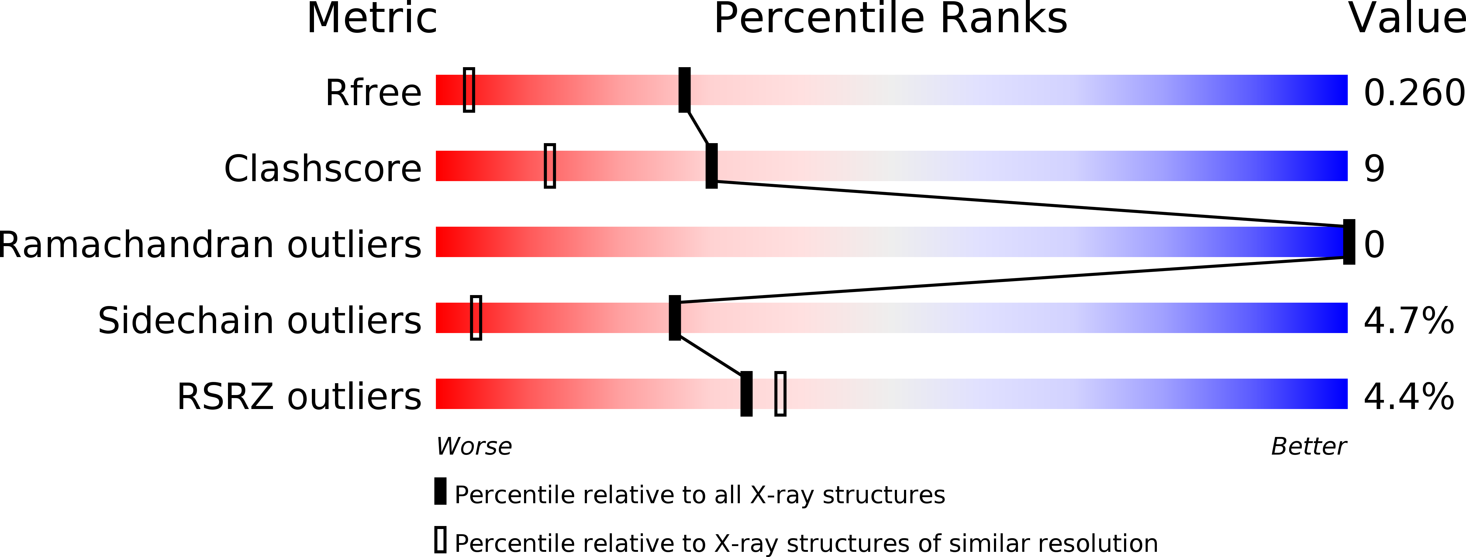
Experimental Data Snapshot
wwPDB Validation 3D Report Full Report
Entity ID: 1 | |||||
|---|---|---|---|---|---|
| Molecule | Chains | Sequence Length | Organism | Details | Image |
| D-3-phosphoglycerate dehydrogenase | A [auth B], B [auth A] | 223 | Homo sapiens | Mutation(s): 0 Gene Names: PHGDH, PGDH3 EC: 1.1.1.95 (PDB Primary Data), 1.1.1.399 (PDB Primary Data), 1.1.1.37 (PDB Primary Data) |  |
UniProt & NIH Common Fund Data Resources | |||||
Find proteins for O43175 (Homo sapiens) Explore O43175 Go to UniProtKB: O43175 | |||||
PHAROS: O43175 GTEx: ENSG00000092621 | |||||
Entity Groups | |||||
| Sequence Clusters | 30% Identity50% Identity70% Identity90% Identity95% Identity100% Identity | ||||
| UniProt Group | O43175 | ||||
Sequence AnnotationsExpand | |||||
| |||||
| Ligands 1 Unique | |||||
|---|---|---|---|---|---|
| ID | Chains | Name / Formula / InChI Key | 2D Diagram | 3D Interactions | |
| 9TT Query on 9TT | C [auth B], D [auth A] | 1-methylindol-5-amine C9 H10 N2 PGTSGPCXPIFQEL-UHFFFAOYSA-N |  | ||
| Length ( Å ) | Angle ( ˚ ) |
|---|---|
| a = 43.342 | α = 97.91 |
| b = 45.86 | β = 110.74 |
| c = 56.175 | γ = 106.39 |
| Software Name | Purpose |
|---|---|
| REFMAC | refinement |
| xia2 | data reduction |
| xia2 | data scaling |
| PHASER | phasing |
| Funding Organization | Location | Grant Number |
|---|---|---|
| Cancer Research UK | United Kingdom | C2115/A21421 |
RCSB PDB (citation) is hosted by
RCSB PDB is a member of the

