5O07

The crystal structure of the human carbonic anhydrase II in complex with a nitroimidazole sulfamate inhibitor


Experimental Data Snapshot

  • Method: X-RAY DIFFRACTION
  • Resolution: 1.80 Å
  • R-Value Free: 0.195 
  • R-Value Work: 0.157 

wwPDB Validation   3D Report Full Report


Ligand Structure Quality Assessment 


This is version 1.2 of the entry. See complete history


Literature

Insights into the binding mode of sulphamates and sulphamides to hCA II: crystallographic studies and binding free energy calculations.

De Simone, G.Langella, E.Esposito, D.Supuran, C.T.Monti, S.M.Winum, J.Y.Alterio, V.

(2017) J Enzyme Inhib Med Chem 32: 1002-1011

  • DOI: https://doi.org/10.1080/14756366.2017.1349764
  • Primary Citation of Related Structures:  
    5O07

  • PubMed Abstract: 

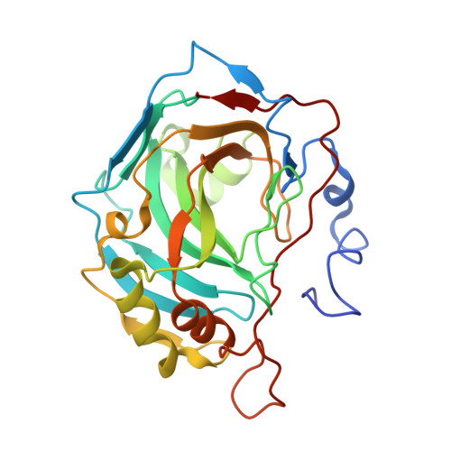
    Sulphamate and sulphamide derivatives have been largely investigated as carbonic anhydrase inhibitors (CAIs) by means of different experimental techniques. However, the structural determinants responsible for their different binding mode to the enzyme active site were not clearly defined so far. In this paper, we report the X-ray crystal structure of hCA II in complex with a sulphamate inhibitor incorporating a nitroimidazole moiety. The comparison with the structure of hCA II in complex with its sulphamide analogue revealed that the two inhibitors adopt a completely different binding mode within the hCA II active site. Starting from these results, we performed a theoretical study on sulphamate and sulphamide derivatives, demonstrating that electrostatic interactions with residues within the enzyme active site play a key role in determining their binding conformation. These findings open new perspectives in the design of effective CAIs using the sulphamate and sulphamide zinc binding groups as lead compounds.


  • Organizational Affiliation

    a Istituto di Biostrutture e Bioimagini , Consiglio Nazionale delle Ricerche , Naples , Italy.


Macromolecules
Find similar proteins by:  (by identity cutoff)  |  3D Structure
Entity ID: 1
MoleculeChains Sequence LengthOrganismDetailsImage
Carbonic anhydrase 2260Homo sapiensMutation(s): 0 
Gene Names: CA2
EC: 4.2.1.1
UniProt & NIH Common Fund Data Resources
Find proteins for P00918 (Homo sapiens)
Explore P00918 
Go to UniProtKB:  P00918
PHAROS:  P00918
GTEx:  ENSG00000104267 
Entity Groups  
Sequence Clusters30% Identity50% Identity70% Identity90% Identity95% Identity100% Identity
UniProt GroupP00918
Sequence Annotations
Expand
  • Reference Sequence
Small Molecules
Ligands 2 Unique
IDChains Name / Formula / InChI Key2D Diagram3D Interactions
1VQ
Query on 1VQ

Download Ideal Coordinates CCD File 
C [auth A]2-(2-methyl-5-nitro-1H-imidazol-1-yl)ethyl sulfamate
C6 H10 N4 O5 S
PBAHHCUVLKOILA-UHFFFAOYSA-N
ZN
Query on ZN

Download Ideal Coordinates CCD File 
B [auth A]ZINC ION
Zn
PTFCDOFLOPIGGS-UHFFFAOYSA-N
Binding Affinity Annotations 
IDSourceBinding Affinity
1VQ Binding MOAD:  5O07 Ki: 33.8 (nM) from 1 assay(s)
BindingDB:  5O07 Ki: 34 (nM) from 1 assay(s)
Experimental Data & Validation

Experimental Data

  • Method: X-RAY DIFFRACTION
  • Resolution: 1.80 Å
  • R-Value Free: 0.195 
  • R-Value Work: 0.157 
  • Space Group: P 1 21 1
Unit Cell:
Length ( Å )Angle ( ˚ )
a = 42.25α = 90
b = 41.252β = 104.26
c = 71.733γ = 90
Software Package:
Software NamePurpose
SCALEPACKdata scaling
CNSrefinement
PDB_EXTRACTdata extraction
HKL-2000data reduction
CrystalCleardata collection
CNSphasing

Structure Validation

View Full Validation Report


Ligand Structure Quality Assessment 


Entry History 

Deposition Data

Revision History  (Full details and data files)

  • Version 1.0: 2017-08-02
    Type: Initial release
  • Version 1.1: 2018-01-24
    Changes: Source and taxonomy
  • Version 1.2: 2024-01-17
    Changes: Data collection, Database references, Refinement description