5NI9

Crystal structure of HLA-DRB1*04:01 with the alpha-enolase peptide 326-340


Experimental Data Snapshot

  • Method: X-RAY DIFFRACTION
  • Resolution: 1.33 Å
  • R-Value Free: 0.176 
  • R-Value Work: 0.146 
  • R-Value Observed: 0.146 

wwPDB Validation   3D Report Full Report


This is version 1.3 of the entry. See complete history


Literature

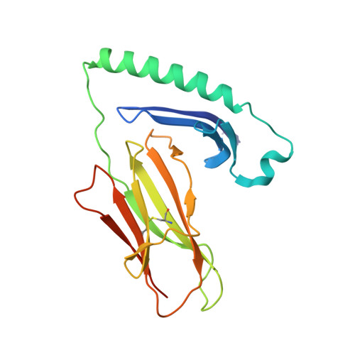
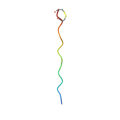
Memory T cells specific to citrullinated alpha-enolase are enriched in the rheumatic joint.

Pieper, J.Dubnovitsky, A.Gerstner, C.James, E.A.Rieck, M.Kozhukh, G.Tandre, K.Pellegrino, S.Gebe, J.A.Ronnblom, L.Sandalova, T.Kwok, W.W.Klareskog, L.Buckner, J.H.Achour, A.Malmstrom, V.

(2018) J Autoimmun 92: 47-56

  • DOI: https://doi.org/10.1016/j.jaut.2018.04.004
  • Primary Citation of Related Structures:  
    5NI9, 5NIG

  • PubMed Abstract: 

    ACPA-positive rheumatoid arthritis (RA) is associated with distinct HLA-DR alleles and immune responses to many citrullinated self-antigens. Herein we investigated the T cell epitope confined within α-enolase 326-340 in the context of HLA-DRB1*04:01 and assessed the corresponding CD4 + T cells in both the circulation and in the rheumatic joint. Comparative crystallographic analyses were performed for the native and citrullinated α-enolase 326-340 peptides in complex with HLA-DRB1*04:01. HLA-tetramers assembled with either the native or citrullinated peptide were used for ex vivo and in vitro assessment of α-enolase-specific T cells in peripheral blood, synovial fluid and synovial tissue by flow cytometry. The native and modified peptides take a completely conserved structural conformation within the peptide-binding cleft of HLA-DRB1*04:01. The citrulline residue-327 was located N-terminally, protruding towards TCRs. The frequencies of T cells recognizing native eno 326-340 were similar in synovial fluid and peripheral blood, while in contrast, the frequency of T cells recognizing cit-eno 326-340 was significantly elevated in synovial fluid compared to peripheral blood (3.6-fold, p = 0.0150). Additionally, citrulline-specific T cells with a memory phenotype were also significantly increased (1.6-fold, p = 0.0052) in synovial fluid compared to peripheral blood. The native T cell epitope confined within α-enolase 326-340 does not appear to lead to complete negative selection of cognate CD4 + T cells. In RA patient samples, only T cells recognizing the citrullinated version of α-enolase 326-340 were found at elevated frequencies implicating that neo-antigen formation is critical for breach of tolerance.


  • Organizational Affiliation

    Rheumatology Unit, Department of Medicine Solna, Center for Molecular Medicine, Karolinska Institutet, Karolinska University Hospital, Stockholm, Sweden.


Macromolecules
Find similar proteins by:  (by identity cutoff)  |  3D Structure
Entity ID: 1
MoleculeChains Sequence LengthOrganismDetailsImage
HLA class II histocompatibility antigen, DR alpha chain189Homo sapiensMutation(s): 0 
Gene Names: HLA-DRAHLA-DRA1
UniProt & NIH Common Fund Data Resources
Find proteins for P01903 (Homo sapiens)
Explore P01903 
Go to UniProtKB:  P01903
PHAROS:  P01903
GTEx:  ENSG00000204287 
Entity Groups  
Sequence Clusters30% Identity50% Identity70% Identity90% Identity95% Identity100% Identity
UniProt GroupP01903
Sequence Annotations
Expand
  • Reference Sequence
Find similar proteins by:  (by identity cutoff)  |  3D Structure
Entity ID: 2
MoleculeChains Sequence LengthOrganismDetailsImage
HLA class II histocompatibility antigen, DRB1-4 beta chain198Homo sapiensMutation(s): 0 
Gene Names: HLA-DRB1
UniProt & NIH Common Fund Data Resources
Find proteins for P01911 (Homo sapiens)
Explore P01911 
Go to UniProtKB:  P01911
PHAROS:  P01911
GTEx:  ENSG00000196126 
Entity Groups  
Sequence Clusters30% Identity50% Identity70% Identity90% Identity95% Identity100% Identity
UniProt GroupP01911
Sequence Annotations
Expand
  • Reference Sequence

Find similar proteins by:  Sequence   |   3D Structure  

Entity ID: 3
MoleculeChains Sequence LengthOrganismDetailsImage
Alpha-enolase15Homo sapiensMutation(s): 0 
EC: 4.2.1.11
UniProt & NIH Common Fund Data Resources
Find proteins for P06733 (Homo sapiens)
Explore P06733 
Go to UniProtKB:  P06733
PHAROS:  P06733
GTEx:  ENSG00000074800 
Entity Groups  
Sequence Clusters30% Identity50% Identity70% Identity90% Identity95% Identity100% Identity
UniProt GroupP06733
Sequence Annotations
Expand
  • Reference Sequence
Experimental Data & Validation

Experimental Data

  • Method: X-RAY DIFFRACTION
  • Resolution: 1.33 Å
  • R-Value Free: 0.176 
  • R-Value Work: 0.146 
  • R-Value Observed: 0.146 
  • Space Group: P 21 21 2
Unit Cell:
Length ( Å )Angle ( ˚ )
a = 68.447α = 90
b = 128.109β = 90
c = 53.585γ = 90
Software Package:
Software NamePurpose
REFMACrefinement
XDSdata reduction
XDSdata scaling
PHASERphasing

Structure Validation

View Full Validation Report


Entry History 

Deposition Data

Revision History  (Full details and data files)

  • Version 1.0: 2018-06-13
    Type: Initial release
  • Version 1.1: 2018-08-01
    Changes: Data collection, Database references
  • Version 1.2: 2019-10-16
    Changes: Data collection
  • Version 1.3: 2024-01-17
    Changes: Data collection, Database references, Refinement description