5NGV

CRYSTAL STRUCTURE OF THE Activin receptor type-2B LIGAND BINDING DOMAIN IN COMPLEX WITH BIMAGRUMAB FV, ORTHORHOMBIC CRYSTAL FORM


Experimental Data Snapshot

  • Method: X-RAY DIFFRACTION
  • Resolution: 2.00 Å
  • R-Value Free: 0.198 
  • R-Value Work: 0.177 
  • R-Value Observed: 0.178 

wwPDB Validation   3D Report Full Report


This is version 2.1 of the entry. See complete history


Literature

Blockade of activin type II receptors with a dual anti-ActRIIA/IIB antibody is critical to promote maximal skeletal muscle hypertrophy.

Morvan, F.Rondeau, J.-M.Zou, C.Minetti, G.Scheufler, C.Scharenberg, M.Jacobi, C.Brebbia, P.Ritter, V.Toussaint, G.Koelbing, C.Leber, X.Schilb, A.Witte, F.Lehmann, S.Koch, E.Geisse, S.Glass, D.J.Lach-Trifilieff, E.

(2017) Proc Natl Acad Sci U S A 114: 12448-12453

  • DOI: https://doi.org/10.1073/pnas.1707925114
  • Primary Citation of Related Structures:  
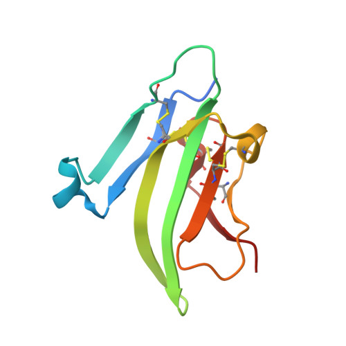
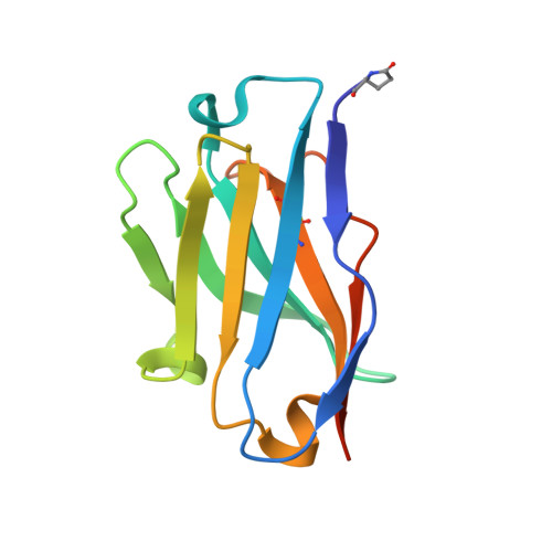
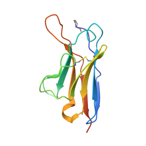
    5NGV, 5NHR, 5NHW

  • PubMed Abstract: 

    The TGF-β family ligands myostatin, GDF11, and activins are negative regulators of skeletal muscle mass, which have been reported to primarily signal via the ActRIIB receptor on skeletal muscle and thereby induce muscle wasting described as cachexia. Use of a soluble ActRIIB-Fc "trap," to block myostatin pathway signaling in normal or cachectic mice leads to hypertrophy or prevention of muscle loss, perhaps suggesting that the ActRIIB receptor is primarily responsible for muscle growth regulation. Genetic evidence demonstrates however that both ActRIIB- and ActRIIA-deficient mice display a hypertrophic phenotype. Here, we describe the mode of action of bimagrumab (BYM338), as a human dual-specific anti-ActRIIA/ActRIIB antibody, at the molecular and cellular levels. As shown by X-ray analysis, bimagrumab binds to both ActRIIA and ActRIIB ligand binding domains in a competitive manner at the critical myostatin/activin binding site, hence preventing signal transduction through either ActRII. Myostatin and the activins are capable of binding to both ActRIIA and ActRIIB, with different affinities. However, blockade of either single receptor through the use of specific anti-ActRIIA or anti-ActRIIB antibodies achieves only a partial signaling blockade upon myostatin or activin A stimulation, and this leads to only a small increase in muscle mass. Complete neutralization and maximal anabolic response are achieved only by simultaneous blockade of both receptors. These findings demonstrate the importance of ActRIIA in addition to ActRIIB in mediating myostatin and activin signaling and highlight the need for blocking both receptors to achieve a strong functional benefit.


  • Organizational Affiliation

    MusculoSkeletal Diseases, Novartis Institutes for Biomedical Research, 4002 Basel, Switzerland.


Macromolecules
Find similar proteins by:  (by identity cutoff)  |  3D Structure
Entity ID: 1
MoleculeChains Sequence LengthOrganismDetailsImage
Activin receptor type-2B99Homo sapiensMutation(s): 0 
Gene Names: ACVR2B
EC: 2.7.11.30
UniProt & NIH Common Fund Data Resources
Find proteins for Q13705 (Homo sapiens)
Explore Q13705 
Go to UniProtKB:  Q13705
PHAROS:  Q13705
GTEx:  ENSG00000114739 
Entity Groups  
Sequence Clusters30% Identity50% Identity70% Identity90% Identity95% Identity100% Identity
UniProt GroupQ13705
Sequence Annotations
Expand
  • Reference Sequence
Find similar proteins by:  (by identity cutoff)  |  3D Structure
Entity ID: 2
MoleculeChains Sequence LengthOrganismDetailsImage
anti-human ActRIIB mAb BYM338 heavy-chainB [auth H]121Homo sapiensMutation(s): 0 
Entity Groups  
Sequence Clusters30% Identity50% Identity70% Identity90% Identity95% Identity100% Identity
Sequence Annotations
Expand
  • Reference Sequence
Find similar proteins by:  (by identity cutoff)  |  3D Structure
Entity ID: 3
MoleculeChains Sequence LengthOrganismDetailsImage
anti-human ActRIIB mAb BYM338 light-chainC [auth L]119Homo sapiensMutation(s): 0 
Entity Groups  
Sequence Clusters30% Identity50% Identity70% Identity90% Identity95% Identity100% Identity
Sequence Annotations
Expand
  • Reference Sequence
Small Molecules
Ligands 1 Unique
IDChains Name / Formula / InChI Key2D Diagram3D Interactions
PG4
Query on PG4

Download Ideal Coordinates CCD File 
D [auth L]TETRAETHYLENE GLYCOL
C8 H18 O5
UWHCKJMYHZGTIT-UHFFFAOYSA-N
Modified Residues  1 Unique
IDChains TypeFormula2D DiagramParent
PCA
Query on PCA
B [auth H]L-PEPTIDE LINKINGC5 H7 N O3GLN
Experimental Data & Validation

Experimental Data

  • Method: X-RAY DIFFRACTION
  • Resolution: 2.00 Å
  • R-Value Free: 0.198 
  • R-Value Work: 0.177 
  • R-Value Observed: 0.178 
  • Space Group: I 2 2 2
Unit Cell:
Length ( Å )Angle ( ˚ )
a = 62.1α = 90
b = 114.17β = 90
c = 117.94γ = 90
Software Package:
Software NamePurpose
XSCALEdata scaling
PHASERphasing
BUSTERrefinement
PDB_EXTRACTdata extraction
XDSdata reduction

Structure Validation

View Full Validation Report


Entry History 

Deposition Data

Revision History  (Full details and data files)

  • Version 1.0: 2017-11-15
    Type: Initial release
  • Version 1.1: 2017-11-29
    Changes: Database references
  • Version 1.2: 2018-01-31
    Changes: Database references, Structure summary
  • Version 2.0: 2020-03-11
    Changes: Polymer sequence
  • Version 2.1: 2024-01-17
    Changes: Data collection, Database references, Refinement description