5MVF

Active structure of EHD4 complexed with ADP


Experimental Data Snapshot

  • Method: X-RAY DIFFRACTION
  • Resolution: 3.27 Å
  • R-Value Free: 0.264 
  • R-Value Work: 0.238 
  • R-Value Observed: 0.239 

wwPDB Validation   3D Report Full Report


Ligand Structure Quality Assessment 


This is version 1.3 of the entry. See complete history


Literature

Structural insights into the activation mechanism of dynamin-like EHD ATPases.

Melo, A.A.Hegde, B.G.Shah, C.Larsson, E.Isas, J.M.Kunz, S.Lundmark, R.Langen, R.Daumke, O.

(2017) Proc Natl Acad Sci U S A 114: 5629-5634

  • DOI: https://doi.org/10.1073/pnas.1614075114
  • Primary Citation of Related Structures:  
    5MTV, 5MVF

  • PubMed Abstract: 

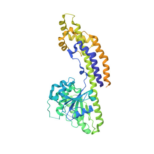
    Eps15 (epidermal growth factor receptor pathway substrate 15)-homology domain containing proteins (EHDs) comprise a family of dynamin-related mechano-chemical ATPases involved in cellular membrane trafficking. Previous studies have revealed the structure of the EHD2 dimer, but the molecular mechanisms of membrane recruitment and assembly have remained obscure. Here, we determined the crystal structure of an amino-terminally truncated EHD4 dimer. Compared with the EHD2 structure, the helical domains are 50° rotated relative to the GTPase domain. Using electron paramagnetic spin resonance (EPR), we show that this rotation aligns the two membrane-binding regions in the helical domain toward the lipid bilayer, allowing membrane interaction. A loop rearrangement in GTPase domain creates a new interface for oligomer formation. Our results suggest that the EHD4 structure represents the active EHD conformation, whereas the EHD2 structure is autoinhibited, and reveal a complex series of domain rearrangements accompanying activation. A comparison with other peripheral membrane proteins elucidates common and specific features of this activation mechanism.


  • Organizational Affiliation

    Crystallography Department, Max-Delbrück-Centrum for Molecular Medicine, 13125 Berlin, Germany.


Macromolecules
Find similar proteins by:  (by identity cutoff)  |  3D Structure
Entity ID: 1
MoleculeChains Sequence LengthOrganismDetailsImage
EH domain-containing protein 4523Mus musculusMutation(s): 0 
Gene Names: Ehd4Past2
UniProt
Find proteins for Q9EQP2 (Mus musculus)
Explore Q9EQP2 
Go to UniProtKB:  Q9EQP2
Entity Groups  
Sequence Clusters30% Identity50% Identity70% Identity90% Identity95% Identity100% Identity
UniProt GroupQ9EQP2
Sequence Annotations
Expand
  • Reference Sequence
Small Molecules
Ligands 2 Unique
IDChains Name / Formula / InChI Key2D Diagram3D Interactions
ADP
Query on ADP

Download Ideal Coordinates CCD File 
B [auth A]ADENOSINE-5'-DIPHOSPHATE
C10 H15 N5 O10 P2
XTWYTFMLZFPYCI-KQYNXXCUSA-N
MG
Query on MG

Download Ideal Coordinates CCD File 
C [auth A]MAGNESIUM ION
Mg
JLVVSXFLKOJNIY-UHFFFAOYSA-N
Experimental Data & Validation

Experimental Data

  • Method: X-RAY DIFFRACTION
  • Resolution: 3.27 Å
  • R-Value Free: 0.264 
  • R-Value Work: 0.238 
  • R-Value Observed: 0.239 
  • Space Group: P 42 21 2
Unit Cell:
Length ( Å )Angle ( ˚ )
a = 199.469α = 90
b = 199.469β = 90
c = 41.79γ = 90
Software Package:
Software NamePurpose
PHENIXrefinement
XDSdata reduction
XSCALEdata scaling
PHASERphasing

Structure Validation

View Full Validation Report


Ligand Structure Quality Assessment 


Entry History & Funding Information

Deposition Data


Funding OrganizationLocationGrant Number
German Research FoundationGermanySFB958/A12

Revision History  (Full details and data files)

  • Version 1.0: 2017-03-08
    Type: Initial release
  • Version 1.1: 2017-06-07
    Changes: Database references
  • Version 1.2: 2017-09-06
    Changes: Author supporting evidence
  • Version 1.3: 2024-01-17
    Changes: Data collection, Database references, Refinement description