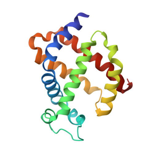
5MJC

metNeuroglobin under oxygen at 50 bar


Experimental Data Snapshot

  • Method: X-RAY DIFFRACTION
  • Resolution: 1.62 Å
  • R-Value Free: 0.207 
  • R-Value Work: 0.168 
  • R-Value Observed: 0.170 

wwPDB Validation   3D Report Full Report


Ligand Structure Quality Assessment 


This is version 1.4 of the entry. See complete history


Literature

Ligand pathways in neuroglobin revealed by low-temperature photodissociation and docking experiments.

Ardiccioni, C.Arcovito, A.Della Longa, S.van der Linden, P.Bourgeois, D.Weik, M.Montemiglio, L.C.Savino, C.Avella, G.Exertier, C.Carpentier, P.Prange, T.Brunori, M.Colloc'h, N.Vallone, B.

(2019) IUCrJ 6: 832-842

  • DOI: https://doi.org/10.1107/S2052252519008157
  • Primary Citation of Related Structures:  
    5MJC, 5MJD, 6I3T, 6I40

  • PubMed Abstract: 

    A combined biophysical approach was applied to map gas-docking sites within murine neuroglobin (Ngb), revealing snapshots of events that might govern activity and dynamics in this unique hexacoordinate globin, which is most likely to be involved in gas-sensing in the central nervous system and for which a precise mechanism of action remains to be elucidated. The application of UV-visible microspectroscopy in crystallo , solution X-ray absorption near-edge spectroscopy and X-ray diffraction experiments at 15-40 K provided the structural characterization of an Ngb photolytic intermediate by cryo-trapping and allowed direct observation of the relocation of carbon monoxide within the distal heme pocket after photodissociation. Moreover, X-ray diffraction at 100 K under a high pressure of dioxygen, a physiological ligand of Ngb, unravelled the existence of a storage site for O 2 in Ngb which coincides with Xe-III, a previously described docking site for xenon or krypton. Notably, no other secondary sites were observed under our experimental conditions.


  • Organizational Affiliation

    Department of Life and Environmental Sciences, New York-Marche Structural Biology Center (NY-MaSBiC), Polytechnic University of Marche, Ancona, Italy.


Macromolecules
Find similar proteins by:  (by identity cutoff)  |  3D Structure
Entity ID: 1
MoleculeChains Sequence LengthOrganismDetailsImage
Neuroglobin148Mus musculusMutation(s): 2 
Gene Names: Ngb
UniProt
Find proteins for Q9ER97 (Mus musculus)
Explore Q9ER97 
Go to UniProtKB:  Q9ER97
Entity Groups  
Sequence Clusters30% Identity50% Identity70% Identity90% Identity95% Identity100% Identity
UniProt GroupQ9ER97
Sequence Annotations
Expand
  • Reference Sequence
Experimental Data & Validation

Experimental Data

  • Method: X-RAY DIFFRACTION
  • Resolution: 1.62 Å
  • R-Value Free: 0.207 
  • R-Value Work: 0.168 
  • R-Value Observed: 0.170 
  • Space Group: H 3 2
Unit Cell:
Length ( Å )Angle ( ˚ )
a = 87.351α = 90
b = 87.351β = 90
c = 112.65γ = 120
Software Package:
Software NamePurpose
REFMACrefinement
XDSdata reduction
XSCALEdata scaling
REFMACphasing

Structure Validation

View Full Validation Report


Ligand Structure Quality Assessment 


Entry History 

Deposition Data

Revision History  (Full details and data files)

  • Version 1.0: 2017-12-20
    Type: Initial release
  • Version 1.1: 2018-02-21
    Changes: Database references
  • Version 1.2: 2019-07-10
    Changes: Data collection, Database references, Derived calculations, Structure summary
  • Version 1.3: 2019-10-16
    Changes: Data collection, Database references
  • Version 1.4: 2024-01-17
    Changes: Data collection, Database references, Refinement description