


Structure of a TRIM21 - UBE2E1 complex reveals the specificity of E2 and ubiquitin recognition by TRIM E3 RINGs
Anandapadamanaban, M., Moche, M., Sunnerhagen, M.To be published.
Experimental Data Snapshot
wwPDB Validation 3D Report Full Report

Entity ID: 1 | |||||
|---|---|---|---|---|---|
| Molecule | Chains | Sequence Length | Organism | Details | Image |
| Ubiquitin-conjugating enzyme E2 E1 | 158 | Homo sapiens | Mutation(s): 0 Gene Names: UBE2E1, UBCH6 EC: 2.3.2.23 (PDB Primary Data), 2.3.2.24 (PDB Primary Data) | 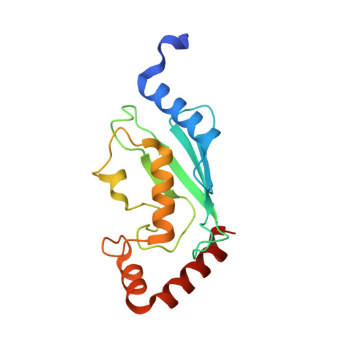 | |
UniProt & NIH Common Fund Data Resources | |||||
Find proteins for P51965 (Homo sapiens) Explore P51965 Go to UniProtKB: P51965 | |||||
PHAROS: P51965 GTEx: ENSG00000170142 | |||||
Entity Groups | |||||
| Sequence Clusters | 30% Identity50% Identity70% Identity90% Identity95% Identity100% Identity | ||||
| UniProt Group | P51965 | ||||
Sequence AnnotationsExpand | |||||
| |||||
| Ligands 1 Unique | |||||
|---|---|---|---|---|---|
| ID | Chains | Name / Formula / InChI Key | 2D Diagram | 3D Interactions | |
| GOL Query on GOL | B [auth A], C [auth A] | GLYCEROL C3 H8 O3 PEDCQBHIVMGVHV-UHFFFAOYSA-N |  | ||
| Length ( Å ) | Angle ( ˚ ) |
|---|---|
| a = 38.301 | α = 90 |
| b = 40.555 | β = 90 |
| c = 91.317 | γ = 90 |
| Software Name | Purpose |
|---|---|
| REFMAC | refinement |
| PDB_EXTRACT | data extraction |
| REFMAC | refinement |
| XDS | data reduction |
| XDS | data scaling |
| PHASER | phasing |
| Funding Organization | Location | Grant Number |
|---|---|---|
| Swedish Research Council | Sweden | -- |
RCSB PDB (citation) is hosted by
RCSB PDB is a member of the

