5LAD

Crystal Structure of apo HydF from thermotoga maritima


Experimental Data Snapshot

  • Method: X-RAY DIFFRACTION
  • Resolution: 3.00 Å
  • R-Value Free: 0.280 
  • R-Value Work: 0.244 
  • R-Value Observed: 0.248 

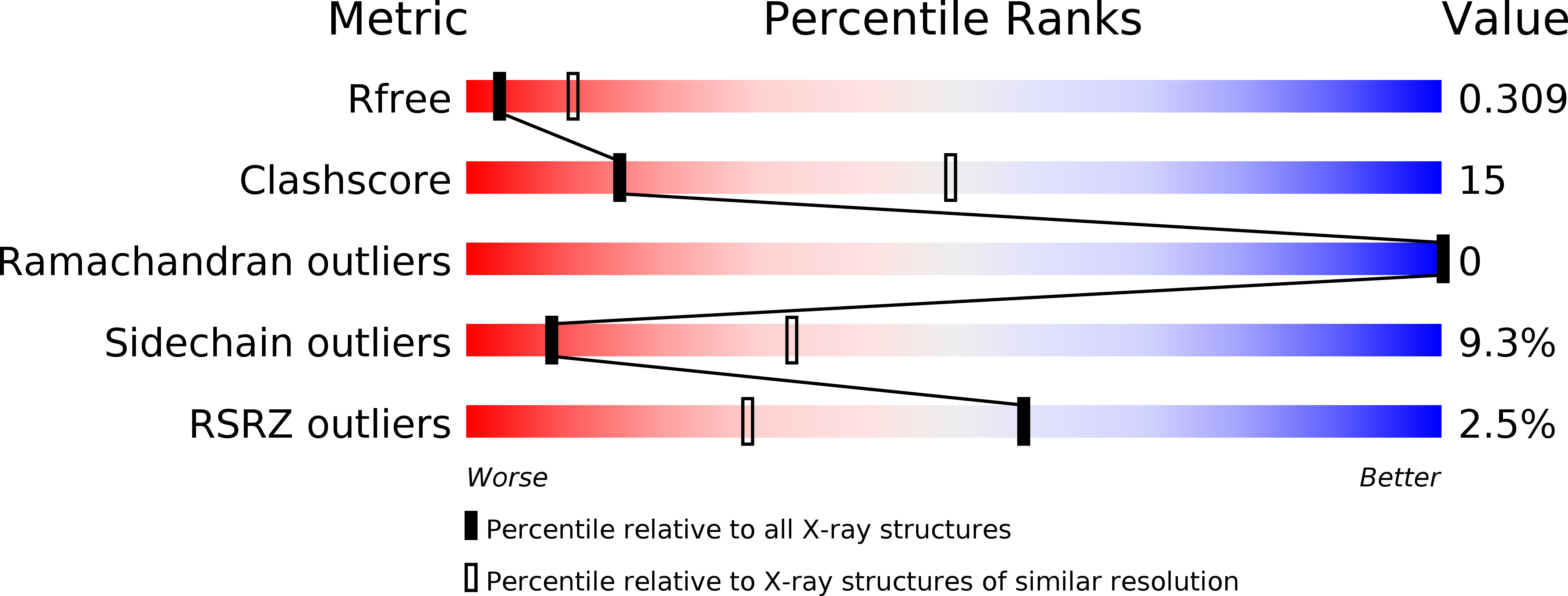
wwPDB Validation   3D Report Full Report


This is version 1.4 of the entry. See complete history


Literature

Structural and functional characterization of the hydrogenase-maturation HydF protein.

Caserta, G.Pecqueur, L.Adamska-Venkatesh, A.Papini, C.Roy, S.Artero, V.Atta, M.Reijerse, E.Lubitz, W.Fontecave, M.

(2017) Nat Chem Biol 13: 779-784

  • DOI: https://doi.org/10.1038/nchembio.2385
  • Primary Citation of Related Structures:  
    5KH0, 5LAD

  • PubMed Abstract: 

    [FeFe] hydrogenase (HydA) catalyzes interconversion between 2H + and H 2 at an active site composed of a [4Fe-4S] cluster linked to a 2Fe subcluster that harbors CO, CN - and azapropanedithiolate (adt 2- ) ligands. HydE, HydG and HydF are the maturases specifically involved in the biosynthesis of the 2Fe subcluster. Using ligands synthesized by HydE and HydG, HydF assembles a di-iron precursor of the 2Fe subcluster and transfers it to HydA for maturation. Here we report the first X-ray structure of HydF with its [4Fe-4S] cluster. The cluster is chelated by three cysteines and an exchangeable glutamate, which allows the binding of synthetic mimics of the 2Fe subcluster. [Fe 2 (adt)(CO) 4 (CN) 2 ] 2- is proposed to be the true di-iron precursor because, when bound to HydF, it matures HydA and displays features in Fourier transform infrared (FTIR) spectra that are similar to those of the native HydF active intermediate. A new route toward the generation of artificial hydrogenases, as combinations of HydF and such biomimetic complexes, is proposed on the basis of the observed hydrogenase activity of chemically modified HydF.


  • Organizational Affiliation

    Laboratoire de Chimie des Processus Biologiques, Collège de France, Université Pierre et Marie Curie, CNRS UMR 8229, Paris, France.


Macromolecules
Find similar proteins by:  (by identity cutoff)  |  3D Structure
Entity ID: 1
MoleculeChains Sequence LengthOrganismDetailsImage
Putative GTP-binding protein404Thermotoga maritimaMutation(s): 0 
Gene Names: TM_0445Tmari_0442
UniProt
Find proteins for Q9WYS6 (Thermotoga maritima (strain ATCC 43589 / DSM 3109 / JCM 10099 / NBRC 100826 / MSB8))
Explore Q9WYS6 
Go to UniProtKB:  Q9WYS6
Entity Groups  
Sequence Clusters30% Identity50% Identity70% Identity90% Identity95% Identity100% Identity
UniProt GroupQ9WYS6
Sequence Annotations
Expand
  • Reference Sequence
Experimental Data & Validation

Experimental Data

  • Method: X-RAY DIFFRACTION
  • Resolution: 3.00 Å
  • R-Value Free: 0.280 
  • R-Value Work: 0.244 
  • R-Value Observed: 0.248 
  • Space Group: I 4
Unit Cell:
Length ( Å )Angle ( ˚ )
a = 126.14α = 90
b = 126.14β = 90
c = 68.84γ = 90
Software Package:
Software NamePurpose
BUSTERrefinement
XDSdata reduction
XDSdata scaling
PHASERphasing

Structure Validation

View Full Validation Report


Entry History 

Deposition Data

Revision History  (Full details and data files)

  • Version 1.0: 2017-05-03
    Type: Initial release
  • Version 1.1: 2017-05-31
    Changes: Database references
  • Version 1.2: 2017-06-07
    Changes: Database references
  • Version 1.3: 2017-06-28
    Changes: Database references
  • Version 1.4: 2024-01-10
    Changes: Data collection, Database references, Refinement description