5K6A

Trypanosoma brucei Pteridine reductase 1 (PTR1) in complex with compound 1


Experimental Data Snapshot

  • Method: X-RAY DIFFRACTION
  • Resolution: 1.70 Å
  • R-Value Free: 0.220 
  • R-Value Work: 0.180 
  • R-Value Observed: 0.182 

wwPDB Validation   3D Report Full Report


Ligand Structure Quality Assessment 


This is version 1.1 of the entry. See complete history


Literature

Chroman-4-One Derivatives Targeting Pteridine Reductase 1 and Showing Anti-Parasitic Activity.

Di Pisa, F.Landi, G.Dello Iacono, L.Pozzi, C.Borsari, C.Ferrari, S.Santucci, M.Santarem, N.Cordeiro-da-Silva, A.Moraes, C.B.Alcantara, L.M.Fontana, V.Freitas-Junior, L.H.Gul, S.Kuzikov, M.Behrens, B.Pohner, I.Wade, R.C.Costi, M.P.Mangani, S.

(2017) Molecules 22

  • DOI: https://doi.org/10.3390/molecules22030426
  • Primary Citation of Related Structures:  
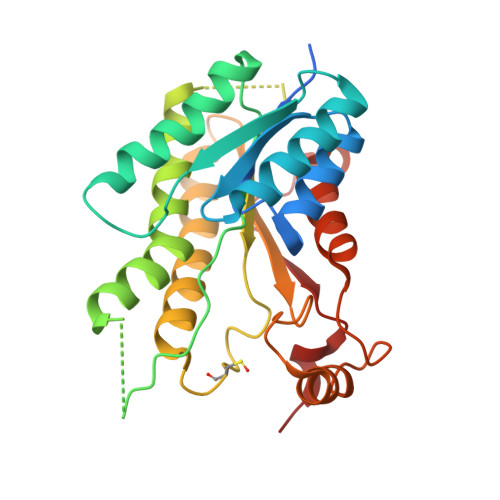
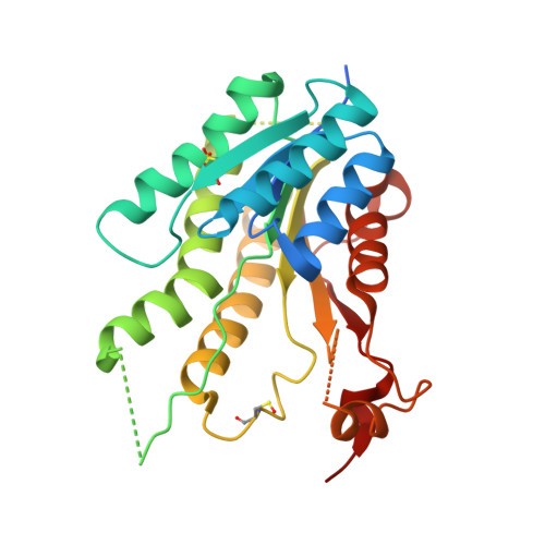
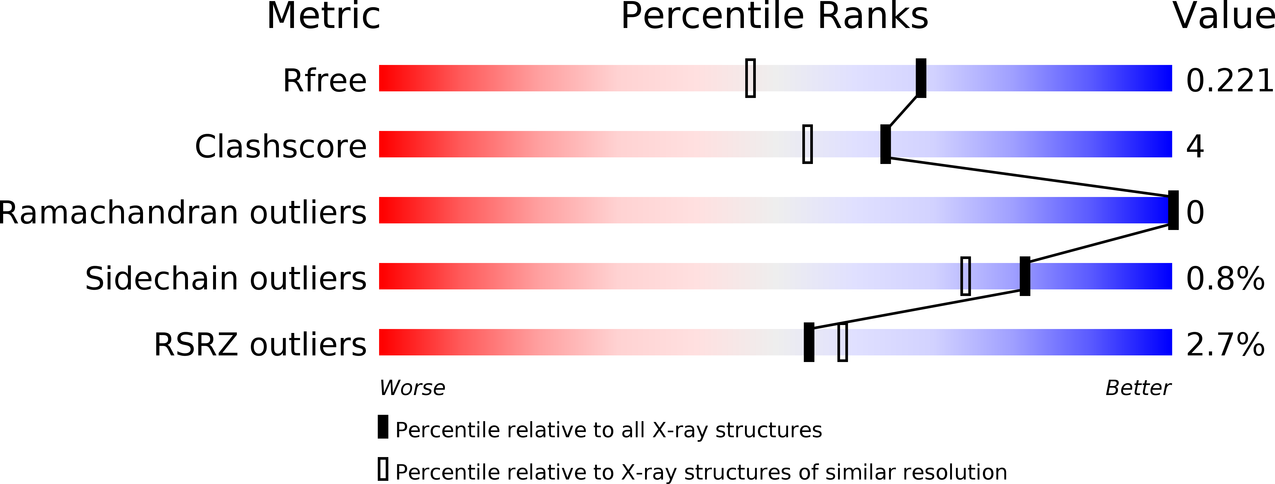
    5K6A, 5L42, 5L4N

  • PubMed Abstract: 

    Flavonoids have previously been identified as antiparasitic agents and pteridine reductase 1 (PTR1) inhibitors. Herein, we focus our attention on the chroman-4-one scaffold. Three chroman-4-one analogues ( 1 - 3 ) of previously published chromen-4-one derivatives were synthesized and biologically evaluated against parasitic enzymes ( Trypanosoma brucei PTR1- Tb PTR1 and Leishmania major-Lm PTR1) and parasites ( Trypanosoma brucei and Leishmania infantum ). A crystal structure of Tb PTR1 in complex with compound 1 and the first crystal structures of Lm PTR1-flavanone complexes (compounds 1 and 3 ) were solved. The inhibitory activity of the chroman-4-one and chromen-4-one derivatives was explained by comparison of observed and predicted binding modes of the compounds. Compound 1 showed activity both against the targeted enzymes and the parasites with a selectivity index greater than 7 and a low toxicity. Our results provide a basis for further scaffold optimization and structure-based drug design aimed at the identification of potent anti-trypanosomatidic compounds targeting multiple PTR1 variants.


  • Organizational Affiliation

    Department of Biotechnology, Chemistry and Pharmacy, University of Siena, 53100 Siena, Italy. dipisa2@unisi.it.


Macromolecules
Find similar proteins by:  (by identity cutoff)  |  3D Structure
Entity ID: 1
MoleculeChains Sequence LengthOrganismDetailsImage
Pteridine reductase
A, B, D
288Trypanosoma brucei bruceiMutation(s): 0 
Gene Names: PTR1
UniProt
Find proteins for O76290 (Trypanosoma brucei brucei)
Explore O76290 
Go to UniProtKB:  O76290
Entity Groups  
Sequence Clusters30% Identity50% Identity70% Identity90% Identity95% Identity100% Identity
UniProt GroupO76290
Sequence Annotations
Expand
  • Reference Sequence
Find similar proteins by:  (by identity cutoff)  |  3D Structure
Entity ID: 2
MoleculeChains Sequence LengthOrganismDetailsImage
Pteridine reductase288Trypanosoma brucei bruceiMutation(s): 0 
Gene Names: PTR1
UniProt
Find proteins for O76290 (Trypanosoma brucei brucei)
Explore O76290 
Go to UniProtKB:  O76290
Entity Groups  
Sequence Clusters30% Identity50% Identity70% Identity90% Identity95% Identity100% Identity
UniProt GroupO76290
Sequence Annotations
Expand
  • Reference Sequence
Small Molecules
Ligands 3 Unique
IDChains Name / Formula / InChI Key2D Diagram3D Interactions
NAP
Query on NAP

Download Ideal Coordinates CCD File 
E [auth A],
H [auth B],
J [auth C],
M [auth D]
NADP NICOTINAMIDE-ADENINE-DINUCLEOTIDE PHOSPHATE
C21 H28 N7 O17 P3
XJLXINKUBYWONI-NNYOXOHSSA-N
6QT
Query on 6QT

Download Ideal Coordinates CCD File 
F [auth A],
I [auth B],
K [auth C],
N [auth D]
(2~{R})-2-(3-hydroxyphenyl)-6-oxidanyl-2,3-dihydrochromen-4-one
C15 H12 O4
FTDCCBIDVVGONG-OAHLLOKOSA-N
ACT
Query on ACT

Download Ideal Coordinates CCD File 
G [auth A],
L [auth C]
ACETATE ION
C2 H3 O2
QTBSBXVTEAMEQO-UHFFFAOYSA-M
Modified Residues  2 Unique
IDChains TypeFormula2D DiagramParent
CSX
Query on CSX
A, B, D
L-PEPTIDE LINKINGC3 H7 N O3 SCYS
OCS
Query on OCS
C
L-PEPTIDE LINKINGC3 H7 N O5 SCYS
Binding Affinity Annotations 
IDSourceBinding Affinity
6QT Binding MOAD:  5K6A IC50: 3.10e+4 (nM) from 1 assay(s)
Experimental Data & Validation

Experimental Data

  • Method: X-RAY DIFFRACTION
  • Resolution: 1.70 Å
  • R-Value Free: 0.220 
  • R-Value Work: 0.180 
  • R-Value Observed: 0.182 
  • Space Group: P 1 21 1
Unit Cell:
Length ( Å )Angle ( ˚ )
a = 74.72α = 90
b = 89.894β = 115.75
c = 82.684γ = 90
Software Package:
Software NamePurpose
REFMACrefinement
iMOSFLMdata reduction
SCALAdata scaling
MOLREPphasing

Structure Validation

View Full Validation Report


Ligand Structure Quality Assessment 


Entry History 

Deposition Data

Revision History  (Full details and data files)

  • Version 1.0: 2017-03-22
    Type: Initial release
  • Version 1.1: 2024-01-10
    Changes: Data collection, Database references, Refinement description